. 2001 Mar 27;98(7):3832-6.
doi: 10.1073/pnas.051515498. Epub 2001 Mar 13.

Profilin I is essential for cell survival and cell division in early mouse development

Affiliations

Profilin I is essential for cell survival and cell division in early mouse development

W Witke et al. Proc Natl Acad Sci U S A. .

Abstract

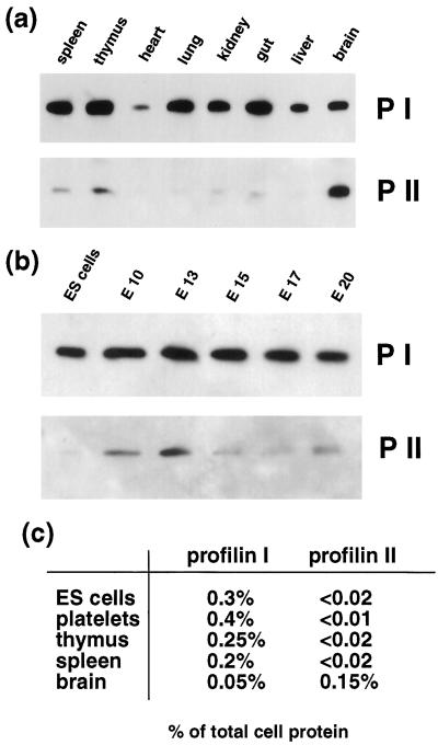
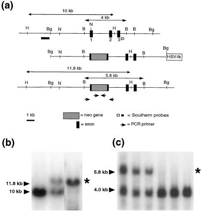
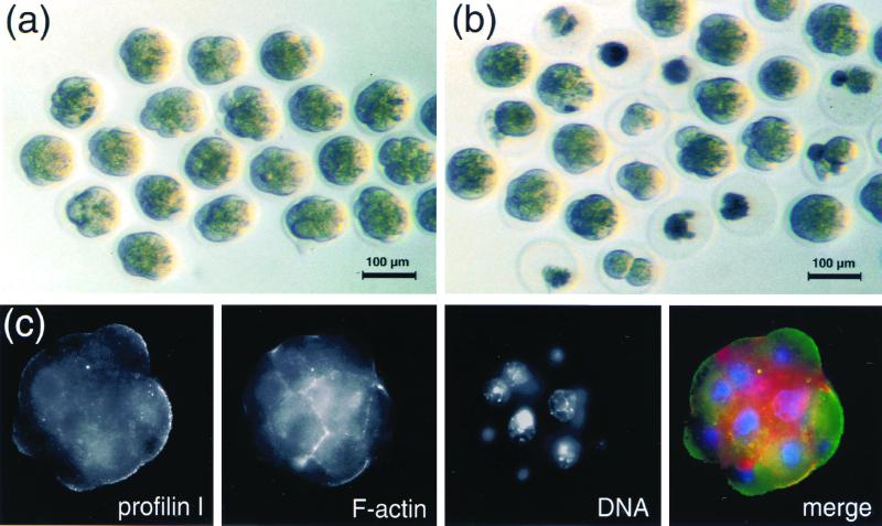
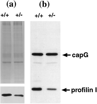
Profilins are thought to play a central role in the regulation of de novo actin assembly by preventing spontaneous actin polymerization through the binding of actin monomers, and the adding of monomeric actin to the barbed actin-filament ends. Other cellular functions of profilin in membrane trafficking and lipid based signaling are also likely. Binding of profilins to signaling molecules such as Arp2/3 complex, Mena, VASP, N-WASP, dynamin I, and others, further implicates profilin and actin as regulators of diverse motile activities. In mouse, two profilins are expressed from two distinct genes. Profilin I is expressed at high levels in all tissues and throughout development, whereas profilin II is expressed in neuronal cells. To examine the function of profilin I in vivo, we generated a null profilin I (pfn1(ko)) allele in mice. Homozygous pfn1(ko/ko) mice are not viable. Pfn1(ko/ko) embryos died as early as the two-cell stage, and no pfn1(ko/ko) blastocysts were detectable. Adult pfn1(ko/wt) mice show a 50% reduction in profilin I expression with no apparent impairment of cell function. However, pfn1(ko/wt) embryos have reduced survival during embryogenesis compared with wild type. Although weakly expressed in early embryos, profilin II cannot compensate for lack of profilin I. Our results indicate that mouse profilin I is an essential protein that has dosage-dependent effects on cell division and survival during embryogenesis.


Figures

Figure 1
Figure 1
Expression of profilin I and profilin IIA protein in mouse tissues and during development. (a) Equal amounts of tissue extracts from adult mice were separated by SDS/PAGE, transferred, and probed with affinity-purified polyclonal antibodies raised against profilin I (P I) and profilin II (P II). (b) Lysates from ES cells and embryos at different stages [embryonic days 10–20 (E10–E20)] were tested for levels of profilins I and IIA. (c) Absolute amounts of profilins I and IIA expressed in various mouse tissues in percentage of total cellular protein.
Figure 2
Figure 2
Generation of profilin I knockout mice. (a) Map of the mouse profilin I gene (Top), the targeting vector (Middle) consisting of an 8.5-kb BglII (Bg) fragment, and the targeted locus (Bottom) are shown. Filled rectangles indicate coding exons 1, 2, and 3. The arrows (Bottom) indicate the primers used to identify the targeted alleles for the PCR analysis of blastocyst DNA. (b) Homologous recombination in ES cells is indicated by generation of an 11.8-kbp fragment (marked with an asterisk) caused by insertion of the neo gene into the wild-type allele of 10 kbp. DNA was tested with the indicated probes (lanes 1 and 2, profilin probe; lane 3, neo probe) after restriction digestion with HindIII (H). (c) Analysis of DNA from litters of a heterozygous breeding. DNA was digested with BamHI and probed with the indicated probe. The targeted allele (5.8 kbp) is marked with an asterisk.
Figure 3
Figure 3
In vitro development of two-cell embryos from wild-type (pfn1wt/wt) and heterozygous (pfn1ko/wt) crossings. Embryos were flushed at E1.5 and cultured for 2 days according to standard procedures (34) to allow development into morulas/blastocysts. (a) Embryos from a pfn1wt/wt mating after 3.5 days. (b) Embryos from a pfn1ko/wt mating after 3.5 days. Note that a significant number of embryos from the mutant cross are growth-arrested and deteriorated. (c) Staining of a wild-type morula for profilin I (green), F-actin (red), and DNA (blue). Embryos were stained with affinity-purified anti-profilin I antibody and an FITC-labeled secondary antibody, tetramethylrhodamine B isothiocyanate-phalloidin for F-actin, and 4′,6-diamidino-2-phenylindole for DNA. Details of the immunostaining protocol are available upon request.
Figure 4
Figure 4
Levels of profilin I in platelets and thymocytes from heterozygous mutant mice. (a) Equal amounts of platelet lysate from pfn1wt/wt and pfn1ko/wt mice were separated by SDS/PAGE, transferred, and probed with a profilin I-specific antibody (Bottom), or the gel was stained with Coomassie blue (Top). The amount of profilin I is reduced by about 50% in the heterozygotes. (b) Equal amounts of thymocyte lysate from pfn1wt/wt and pfn1ko/wt mice were subjected to SDS/PAGE and probed with a profilin I-specific antibody. The filter was reprobed with a (capG)-specific antibody to normalize for loaded protein amounts. Note the reduction of profilin I in the mutant thymocytes.

References

    1. Carlsson L, Nystrom L E, Sundkvist I, Markey F, Lindberg U. J Mol Biol. 1977;115:465–483. - PubMed
    1. Reichstein E, Korn E D. J Biol Chem. 1979;254:6174–6179. - PubMed
    1. Valenta R, Duchene M, Ebner C, Valent P, Sillaber C, Deviller P, Ferreira F, Tejkl M, Edelmann H, Kraft D, et al. J Exp Med. 1992;175:377–385. - PMC - PubMed
    1. Machesky L M, Cole N B, Moss B, Pollard T D. Biochemistry. 1994;33:10815–10824. - PubMed
    1. Goldschmidt-Clermont P J, Furman M I, Wachsstock D, Safer D, Nachmias V T, Pollard T D. Mol Biol Cell. 1992;3:1015–1024. - PMC - PubMed

Publication types