. 2001 Mar 27;98(7):3855-60.
doi: 10.1073/pnas.071586898.

A splicing switch and gain-of-function mutation in FgfR2-IIIc hemizygotes causes Apert/Pfeiffer-syndrome-like phenotypes

Affiliations

A splicing switch and gain-of-function mutation in FgfR2-IIIc hemizygotes causes Apert/Pfeiffer-syndrome-like phenotypes

M K Hajihosseini et al. Proc Natl Acad Sci U S A. .

Abstract

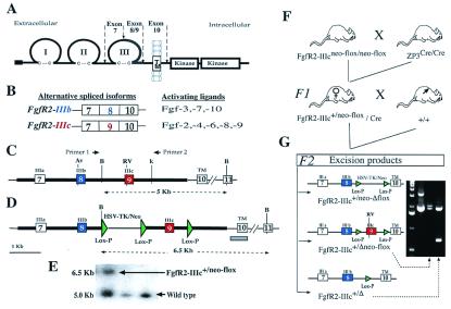
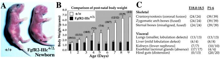
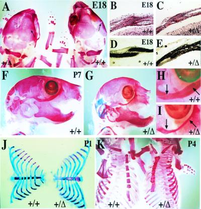
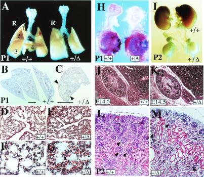
Intercellular signaling by fibroblast growth factors plays vital roles during embryogenesis. Mice deficient for fibroblast growth factor receptors (FgfRs) show abnormalities in early gastrulation and implantation, disruptions in epithelial-mesenchymal interactions, as well as profound defects in membranous and endochondrial bone formation. Activating FGFR mutations are the underlying cause of several craniosynostoses and dwarfism syndromes in humans. Here we show that a heterozygotic abrogation of FgfR2-exon 9 (IIIc) in mice causes a splicing switch, resulting in a gain-of-function mutation. The consequences are neonatal growth retardation and death, coronal synostosis, ocular proptosis, precocious sternal fusion, and abnormalities in secondary branching in several organs that undergo branching morphogenesis. This phenotype has strong parallels to some Apert's and Pfeiffer's syndrome patients.
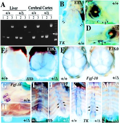

Figures

Figure 1
Figure 1
Fgf receptor structure and strategy for deletion of FgfR2 exon 9 (IIIc). (A) Schematic structure of FgfR2 highlighting the third Ig loop, a region in which alternative usage of exons 8 or 9 in FgfR2 leads to the generation of the IIIb or IIIc isoforms, respectively. TM, transmembrane domain. (B) The main ligands activating each of these isoforms. (C) Schematic depiction of mouse genomic DNA encompassing FgfR2 exons (boxes) 7, 8, 9, and 10, drawn to scale and showing main restriction enzyme sites (Av, AvaI; RV, EcoRV; B, BamHI; K, KpnI). (D) Targeting construct showing loxP sequences placed downstream of exon 9, and flanking the selectable marker gene neo driven by HSV-TK promoter located upstream of exon 9. Thick lines indicate the extent of the targeting construct. (E) Homologous recombinant 129 embryonic stem cells were identified by Southern blotting of BamHI-digested DNA by using a 450-bp genomic probe located 3′ of target vector sequences (gray bar in D). Homologous recombinant cells (FgfR2-IIIc+/neo-flox) yielded a 6.5-kb fragment and wild type yielded a 5.0-kb fragment. Both 5′ and 3′ joins were checked by PCR analysis. (F) Hemizygous (FgfR2-IIIc+/Δ) mutant mice were generated by crossing FgfR2-IIIcneo-flox/neo-flox with ZP3-Cre females (34), and then crossing F1 females carrying one copy of the targeting construct as well as the Cre transgene with wild-type males. Approximately 50% of such females gave Cre-mediated excision. (G) Of these excisions, 90% were complete (FgfR2-IIIc+/Δ); the remainder excised only the selectable marker (FgfR2-IIIc+/Δneo-flox), as determined by PCR using the pair of primers shown in C. The potential excision product, FgfR2-IIIc+/neo-Δflox, was not observed.
Figure 2
Figure 2
Growth retardation in FgfR2-IIIc+/Δ mice. (A) Hemizygotes were identifiable at birth by their smaller sizes, characteristic dome-shaped heads, and truncated faces; the genotype was confirmed by PCR using tail genomic DNA (Fig. 1G). (B) All hemizygotes showed growth retardation after birth as reflected in a comparison of their body weights with those of wild-type littermates. Brackets show the number of hemizygotes and wild types analyzed. (C) Table summarizing the major abnormalities and their penetrance (number showing specified defects/total number of hemizygotes analyzed) at E18 and postnatally.
Figure 3
Figure 3
Precocious ossification of coronal sutures and the sternum in FgfR2-IIIc+/Δ mice. Skeletons stained with alizarin red to identify ossified tissue (A and K), or stained with alizarin red in combination with Alcian blue stain to additionally reveal cartilage (FJ). (A) Dorsal view of calvarial bones at E18 showing closer apposition of frontal and parietal bones at the coronal suture (arrows) in a hemizygote compared with its wild-type litter mate. (BE) Transverse sections through the frontal and parietal bones showing fusion of coronal sutures in hemizygote in contrast to wild-type mice. Sections stained with hematoxylin–eosin (B and C) or alkaline phosphatase (D and E). (FI) Lateral views of skulls from 7-day-old mice showing in hemizygotes rounded heads, truncated maxilla, and fusion of joints separating the zygomatic arch bones (part of the maxilla, zygomatic, and temporal bones) which make up the lower rim of the eye socket. (H and I) Detail of zygomatic arch joints shown in F and G, respectively. (J and K) Dissected and whole rib cages from 1- and 4-day-old mice, respectively, showing precocious and progressive sternal fusion in hemizygotes. Note that in these mice, individual sternebrae are thicker and less congruent, and the manubrium and xiphoid processes are bifurcated.
Figure 4
Figure 4
Visceral defects in FgfR2-IIIc+/Δ mice. (A) Comparison of whole lungs at postnatal day 1 (P1) showing the development of a single right lobe in hemizygotes compared with four distinct lobes in the wild type. (BG) Transverse sections through the right lobes stained with hematoxylin–eosin showing partial lobe separation in the mutant right lung (indicated by arrowheads in C), as well as the development of fewer bronchioles lined with ciliated cells and fewer branched alveolar structures (D and E). (B and C, bars = 4 mm.) In mutants, lung mesenchyme is more compact and often congested with red blood cells (F and G). (H) Dissected exorbital lacrimal glands with their surrounding mesenchyme still attached to the eye and stained with the dye carmalum to reveal branching, show a clear lack of gland development in hemizygotes. (I) Comparison of kidneys at P2 reveals severe growth retardation in hemizygotes which does not seem to affect the adrenal glands lying above each kidney. (J and K) Saggital hematoxylin–eosin (H&E)-stained sections through E14.5 embryonic kidneys show the presence of fewer developing nephrons in the cortical region of hemizygotes. (L and M) H&E-stained transverse section of P2 kidneys, showing fewer and degenerating glomeruli (arrowheads), dilated proximal and distal tubules, and more undifferentiated mesenchyme in hemizygotes.
Figure 5
Figure 5
Analysis of FgfR2 and Fgf-10 expression in brain, calvaria, lacrimal glands, and sternum. (A) Reverse transcription–PCR assessment of FgfR2 expression in the liver and cerebral cortices (dissected free of meninges) of wild-type and hemizygous mice by using primers from within exons 7 and 8 [IIIb specific (320 bp), lane 1], or exons 7 and 9 [IIIc specific (310 bp), lane 2], or exons 7 and 10 [detects both isoforms (495 and 489 bp), lane 3]. Identity of bands was confirmed by DNA sequencing. Note that the IIIb-specific product, as well as a faint 345-bp band (asterisks), derived from IIIa-TM splice, is detected in the cerebral cortex and liver of hemizygous but not wild-type mice. (BD) Expression pattern of FgfR2 in E17.5 calvaria of wild type (+/+) and hemizygote (+/Δ) as revealed by whole mount in situ hybridization (WMISH) using digoxigenin-labeled TK probe which is isoform nonspecific. Arrows in B point to staining of coronal sutures of wild type. Staining was less intense in hemizygotes (data not shown). (C and D) Bones making up the lower rim of the eye socket, the zygomatic arch (arrowheads), as well as their intervening joints are also labeled; these joints (double arrowheads) are more closely apposed in hemizygotes than in wild types. (E) WMISH with an FgfR2-IIIb-specific probe on E18.5 calvaria shows elevated expression of this isoform in calvarial sutures of hemizygotes but not in wild-type mice. Hemizygotes also present a shortened snout, wider skull, and delayed closure of the anterior fontanelle. (F) WMISH with an Fgf-10 probe shows expression of this ligand in the suture regions of both wild types and hemizygotes at E18. (G and H) In the same specimens shown in F, Fgf-10 probe labels the tips of branches within the exorbital lacrimal glands of wild type (G), whereas there is a lack of expression in hemizygote gland rudiment (H). (IM) WMISH analysis on dissected sternums from E18 mice with an FgfR2-IIIb-specific probe (I and J), E18.5 mice using a TK probe (K and L), and E18 using an Fgf-10 probe (M). In both wild types and hemizygotes, TK and IIIb strongly label the periosteal regions of each sternebrae. However, delayed midline fusion results in two hemi sternebrae in hemizygotes (arrows in J and L), the medial rims of which also express TK and IIIb. At E18.5, when medial fusion is more advanced, TK additionally labels the periphery of the xiphoid processes, which appears bifurcated in hemizygotes. (M) Fgf-10 is expressed by interrib periosteal regions as well as the body of xiphoid.

Comment in

References

    1. McKeehan W L, Wang F, Kan M. Prog Nucleic Acid Res Mol Biol. 1998;59:135–176. - PubMed
    1. Ornitz D M. BioEssays. 2000;22:108–112. - PubMed
    1. Nishimura T, Nakatake Y, Konishi M, Itoh N. Biochem Biophys Acta. 2000;1492:203–206. - PubMed
    1. Yamashita T, Yoshioka M, Itoh N. Biochem Biophys Res Commun. 2000;277:494–498. - PubMed
    1. Ornitz D M, Xu J S, Colvin J S, McEwen D G, MacArthur C A, Coulier F, Gao G X, Goldfarb M. J Biol Chem. 1996;271:15292–15297. - PubMed

Publication types

MeSH terms

Substances

LinkOut - more resources