. 2001 Mar 27;98(7):4060-5.
doi: 10.1073/pnas.071594398.

A serologically identified tumor antigen encoded by a homeobox gene promotes growth of ovarian epithelial cells

Affiliations

A serologically identified tumor antigen encoded by a homeobox gene promotes growth of ovarian epithelial cells

H Naora et al. Proc Natl Acad Sci U S A. .

Abstract

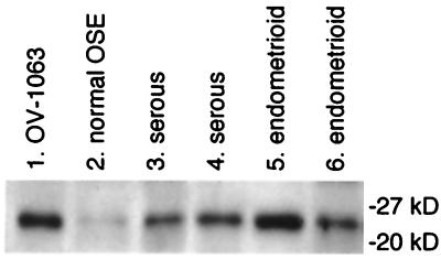
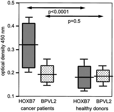
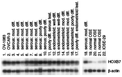
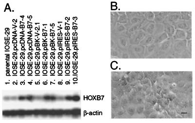
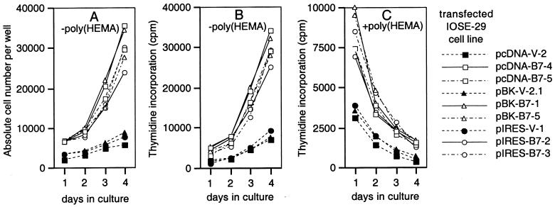
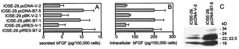
Ovarian carcinomas are thought to arise from cells of the ovarian surface epithelium by mechanisms that are poorly understood. Molecules associated with neoplasia are potentially immunogenic, but few ovarian tumor antigens have been identified. Because ovarian carcinomas can elicit humoral responses in patients, we searched for novel tumor antigens by immunoscreening a cDNA expression library with ovarian cancer patient serum. Seven clones corresponding to the homeobox gene HOXB7 were isolated. ELISAs using purified recombinant HOXB7 protein revealed significant serologic reactivity to HOXB7 in 13 of 39 ovarian cancer patients and in only one of 29 healthy women (P < 0.0001). Ovarian carcinomas were found to express HOXB7 at markedly higher levels than normal ovarian surface epithelium, suggesting that immunogenicity of HOXB7 in patients could be associated with its elevated expression in ovarian carcinomas. Overexpression of HOXB7 in immortalized normal ovarian surface epithelial cells dramatically enhanced cellular proliferation. Furthermore, HOXB7 overexpression increased intracellular accumulation and secretion of basic fibroblast growth factor, a potent angiogenic and mitogenic factor. These results reveal HOXB7 as a tumor antigen whose up-regulated expression could play a significant role in promoting growth and development of ovarian carcinomas.


Figures

Figure 1
Figure 1
Detection of a protein common to OV-1063 cells and ovarian carcinomas by Western blot analysis by using patient serum. Protein lysates (10 μg per lane) prepared from OV-1063 cells (lane 1), normal OSE (lane 2), and ovarian carcinomas of the serous (lanes 3 and 4) and endometrioid (lanes 5 and 6) histotypes were probed with diluted serum (1:500) of a patient with Stage III serous ovarian carcinoma. Shown are bands corresponding to a common reactive species. Positions of protein size markers are indicated.
Figure 2
Figure 2
Serologic responses to HOXB7 as assessed by ELISA. BPVL2 protein was used as a negative control protein. Sera were diluted 1:500. Shown are values of statistical significance for differences in optical density at 450 nm of sera of patients (n = 39) and of healthy women (n = 29) as determined by the Mann–Whitney u test. Statistical significance for differences in responses to HOXB7 and to BPVL2 were assessed by the Wilcoxon's signed-rank test for paired data and were found to be P < 0.0001 for patients and P = 0.4 for healthy women. Horizontal bars indicate median values
Figure 3
Figure 3
Semiquantitative RT-PCR analysis of HOXB7 expression. Shown are Southern blots of HOXB7 and β-actin RT-PCR products in OV-1063, OVCAR-3 and IOSE-29 cells (lanes 1, 2, and 22), specimens of normal OSE (lanes 19–21) and ovarian carcinomas (lanes 3–18). Histology of carcinomas ranged from poorly differentiated (diff.) with either serous or endometrioid features (feat.) to moderately (mod.) and well-differentiated serous and endometrioid. The specimen used for analysis shown in lane 18 is the same as that in lane 6.
Figure 4
Figure 4
HOXB7 expression levels and morphology of transfected IOSE-29 cells. (A) RT-PCR analysis detected HOXB7 expression levels in HOXB7-transfected cells (lanes 3, 4, 6, 7, 9, 10) that were markedly higher than in cells transfected with vector DNA alone (lanes 2, 5, 8) and in the parental cell line (lane 1). Phase-contrast microscopy revealed that IOSE-29 cells transfected with vector DNA grew in flat monolayers (B), whereas cultures of HOXB7-transfected cells exhibited islands of multilayered overgrowth (C). (Bar = 10 μm.)
Figure 5
Figure 5
Growth characteristics of transfected cells. Vector- and HOXB7-transfected IOSE-29 cells were seeded at 2,000 cells per well in uncoated 96-well plates (A and B) and wells coated with poly(HEMA) (C). Total numbers of cells in each uncoated well were counted daily (A). After 1, 2, 3, and 4 days in culture, thymidine incorporation was measured in cells pulsed with [3H]methylthymidine for 3 h in uncoated wells (B), and for 18 h in poly(HEMA)-coated wells (C). Shown are the mean values of three to four independent experiments. Differences in cell numbers and thymidine incorporation levels of HOXB7-transfected cells, as compared with corresponding vector-transfected cells, at each time point were found to be statistically significant (P < 0.001).
Figure 6
Figure 6
Increased bFGF production by HOXB7-transfected cells. bFGF levels in (A) culture supernatants collected from equivalent numbers of vector- and HOXB7-transfected IOSE-29 cells and (B) lysates of these cells were assessed by ELISA. Shown are mean values of two to three independent experiments. (C) Western blot analysis of lysates (10 μg per lane) of vector-transfected (IOSE-29.pcDNA-V-2) and HOXB7-transfected (IOSE-29.pcDNA-B7–5) IOSE-29 cells. Molecular weights of bFGF isoforms are indicated.

References

    1. Rosenthal A, Jacobs I. Semin Oncol. 1998;25:315–325. - PubMed
    1. Taylor D D, Gercel-Taylor C. Oncol Rep. 1998;5:1519–1524. - PubMed
    1. Slamon D J, Godolphin W, Jones L A, Holt J A, Wong S G, Keith D E, Levin W J, Stuart S G, Udove J, Ullrich A, Press M F. Science. 1989;24:707–712. - PubMed
    1. Chen Y-T, Scanlan M J, Sahin U, Tureci O, Gure A O, Tsang S, Williamson B, Stockert E, Pfreundschuh M, Old L J. Proc Natl Acad Sci USA. 1997;94:1914–1918. - PMC - PubMed
    1. Russo V, Dalerba P, Ricci A, Bonazzi C, Leone B E, Mangioni C, Allavera P, Bordignon C, Traversari C. Int J Cancer. 1996;67:457–460. - PubMed

Publication types

MeSH terms