. 2001 Mar 27;98(7):4078-83.
doi: 10.1073/pnas.061004098.

Local gene transfer of tissue factor pathway inhibitor regulates intimal hyperplasia in atherosclerotic arteries

Affiliations

Local gene transfer of tissue factor pathway inhibitor regulates intimal hyperplasia in atherosclerotic arteries

P Zoldhelyi et al. Proc Natl Acad Sci U S A. .

Abstract

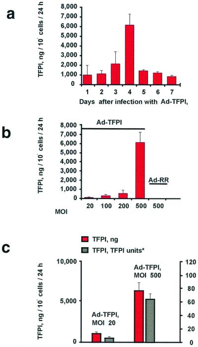
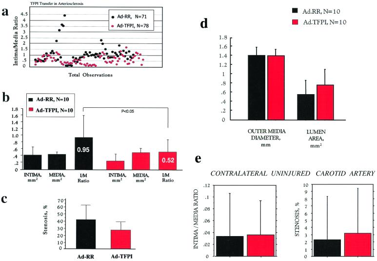
Tissue factor (TF), the initiator of blood coagulation and thrombosis, is up-regulated after vascular injury and in atherosclerotic states. Systemic administration of recombinant TF pathway inhibitor (TFPI) has been reported to decrease intimal hyperplasia after vascular injury and also to suppress systemic mechanisms of blood coagulation and thrombosis. Here we report that, in heritable hyperlipidemic Watanabe rabbits, adenoviral gene transfer of TFPI to balloon-injured atherosclerotic arteries reduced the extent of intimal hyperplasia by 43% (P < 0.05) compared with a control vector used at identical titer (1 x 10(10) plaque-forming units/ml). Platelet aggregation and coagulation studies performed 7 days after local gene transfer of TFPI failed to show any impairment in systemic hemostasis. At time of sacrifice, 4 weeks after vascular injury, the 10 Ad-TFPI treated carotid arteries were free of thrombi, whereas two control-treated arteries were occluded (P, not significant). These findings suggest that TFPI overexpressed in atherosclerotic arteries can regulate hyperplastic response to injury in the absence of changes in the hemostatic system, establishing a role for local TF regulation as target for gene transfer-based antirestenosis therapies.


Figures

Figure 1
Figure 1
TFPI secretion in canine VSMC. Human TFPI was measured by the ELISA in the conditioned medium of dog VSMC. (a) Time course of TFPI (in ng/106 cells/24 h) secreted by VSMC infected for 6 h with Ad-TFPI or Ad-RR (identical construct minus the foreign gene) at moi 500 (see Methods). (b) Viral dose response of TFPI secretion in VSMC (ng/106 cells/24 h) 4 days after a 6-h infection with Ad-TFPI. No TFPI was detected in VSMC infected with Ad-RR. (c) Anti-TF/factor VIIa activity of human TFPI secreted by dog VSMC. TFPI activity in the conditioned medium is expressed in TFPI units (where 1 unit corresponds to about 55 ng TFPI in human plasma; see Methods).
Figure 2
Figure 2
TFPI gene transfer in balloon-injured atherosclerotic rabbit arteries and TFPI immunoreactivity in normal rabbit endothelium. Atherosclerotic carotid plaque of a Watanabe rabbit killed 4 days after infection with Ad-TFPI at 1 × 10¹⁰ pfu/ml (see Methods). (a) Immunostain for human TFPI. (b) The TFPI antibody used in a is replaced with an unrelated (cytomegalovirus) control antibody in an adjacent section; c and d show endothelium, respectively, of the carotid artery and s.c. microvasculature of a normal New Zealand White rabbit killed without prior injury. Tissue sections were immunostained with the antibody recognizing human TFPI. a and b, ×200; c, ×400; d, ×100.
Figure 3
Figure 3
Histomorphometric observations in balloon-injured carotid arteries of Watanabe rabbits treated with Ad-TFPI. The carotid arteries of 12- to 16-month-old Watanabe rabbits of either sex were treated at the time of severe balloon injury with Ad-TFPI, encoding a human TFPI cDNA, or Ad-RR, an identical vector minus the transgene. Both vectors were locally delivered at a titer of 1 × 10¹⁰ pfu/ml. Animals were killed 4 weeks after injury, and carotid arteries were processed as described in Methods. Quantitative histomorphometry was performed in blinded fashion. Shown are (a) individual intima/media ratios observed in injured and adenovirally treated arterial sections; (b) mean areas of intima and media and mean intima/media ratios; (c) the degree of stenosis in the injured arteries; (d) the average outer diameter of the media, computed as average diameter between opposite sides of the media/adventitia border (a measure of external vessel size independent of dissection artifacts of the adventitia) and the average lumen area; and (e) intima/media ratio and stenosis of the uninjured contralateral carotid arteries.
Figure 4
Figure 4
Examples of carotid arteries of Watanabe rabbits. (a and b) Verhoeft-Van Gieson stain of sections from Watanabe rabbit carotid arteries treated with Ad-TFPI (a) and Ad-RR (b). (c) Plaque in the noninjured contralateral carotid artery of an Ad-TFPI-treated rabbit. (d) Section adjacent to that shown in c and stained with hematoxylin-eosin to illustrate the difference in texture between arterial tissue and latex filled arterial lumen. I, intima; L, lumen filled with residual latex to prevent postmortem distortion of lumen and vessel wall; P, atherosclerotic plaque. →, internal elastic membrane. (×40.)

References

    1. Lefkovits J, Topol E. Prog Cardiovasc Dis. 1997;40:141–158. - PubMed
    1. Schwartz S. Circ Res. 1999;85:877–879. - PubMed
    1. Willerson J, Yao S, McNatt J, Benedict C, Anderson H, Golino P, Murphree S, Buja L. Proc Natl Acad Sci USA. 1991;88:10624–10628. - PMC - PubMed
    1. Sato Y, Asada Y, Marutsuka K, Hatakeyama K, Kamikubo Y, Sumiyoshi A. Thromb Haemostasis. 1997;78:1138–1141. - PubMed
    1. Feng N, Yang Y, Huang S, Ou J. J Clin Invest. 1996;98:1493–1501. - PMC - PubMed

Publication types

Substances

LinkOut - more resources