. 2001 Mar 27;98(7):4113-8.
doi: 10.1073/pnas.071432398.

Modular organization of the Friend murine leukemia virus envelope protein underlies the mechanism of infection

Affiliations

Modular organization of the Friend murine leukemia virus envelope protein underlies the mechanism of infection

A L Barnett et al. Proc Natl Acad Sci U S A. .

Abstract

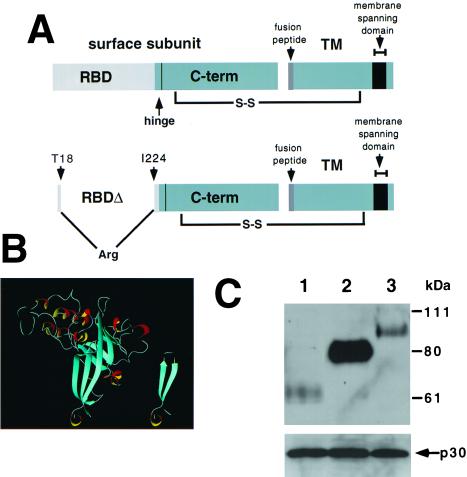
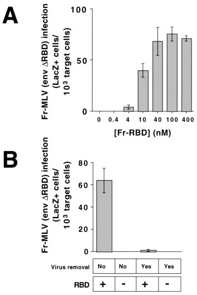
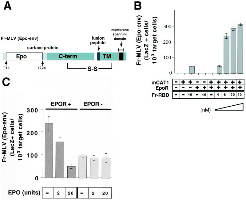
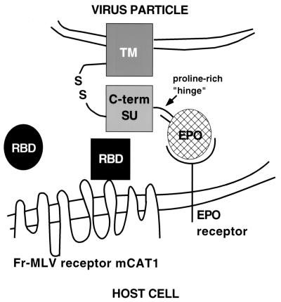
Retrovirus infection is initiated by receptor-dependent fusion of the envelope to the cell membrane. The modular organization of the envelope protein of C type retroviruses has been exploited to investigate how binding of the surface subunit (SU) to receptor triggers fusion mediated by the transmembrane (TM) subunit. We show that deletion of the receptor-binding domain (RBD) from SU of Friend murine leukemia virus (Fr-MLV) abolishes infection that is restored by supplying RBD as a soluble protein. Infection by this mechanism remains dependent on receptor expression. When membrane attachment of the virus lacking RBD is reestablished by inserting the hormone erythropoietin, infection remains dependent on the RBD/receptor complex. However, infection increases 50-fold to 5 x 10(5) units/ml on cells that also express the erythropoietin receptor. Soluble RBD from Fr-MLV also restores infection by amphotropic and xenotropic MLVs in which RBD is deleted. These experiments demonstrate that RBD has two functions: mediating virus attachment and activating the fusion mechanism. In addition, they indicate that receptor engagement triggers fusion by promoting a subgroup-independent functional interaction between RBD and the remainder of SU and/or TM.


Figures

Figure 1
Figure 1
(A) Schematic diagram of RBD in Fr-MLV envelope glycoprotein. The top diagram illustrates that RBD is composed of the N-terminal portion of the SU and is connected to the C-terminal portion of SU by a proline-rich “hinge.” The SU and TM are linked by a disulfide bond. The bottom diagram illustrates the location of the deletion (residues 19–223) and insertion of an arginine residue introduced to obtain Fr-MLV (env ΔRBD). (B) Proposed structure of Fr-MLV Env ΔRBD. Ribbon diagram of the RBD of the Fr-MLV surface glycoprotein (Left). It is composed of a barrel-like structure (blue) similar to an Ig-fold and a series of loops (white) and helices (red/yellow) at the top that contact receptor (10, 11). The N- and C-terminal β-strands are adjacent and together form the base of RBD. Env ΔRBD was prepared by deleting residues 19–223 and connecting these two β-strands by inserting an arginine residue between T18 and I224 at the top of the stalk. The env ΔRBD structure was modeled in the Swiss Protein Data Bank, selecting a structure that minimized steric clashes, and was visualized by using molmol (Right). (C) Incorporation of MLV envelope proteins into virions. Immunoblot of a nitrocellulose filter prepared from lysates of purified virions after SDS/PAGE. The top filter was probed with goat anti-MLV gp70 and HRP mouse anti-goat antibodies. Lane 1: lysate from Fr-MLV (env ΔRBD). Lane 2: lysate from wild-type Fr-MLV. Lane 3: lysate from Fr-MLV (Epo-env). To control for MLV particle production, aliquots of the same lysates were examined for the presence of the p30 capsid protein after SDS/PAGE by using goat anti-MLV and HRP mouse anti-goat antibodies (below).
Figure 2
Figure 2
Rescue of Fr-MLV (env ΔRBD) infection by soluble RBD on 293 mCAT1 cells. (A) In each well of a 6-well plate, 5 × 10⁵ human 293-derived cells expressing the Fr-MLV receptor, mCAT1, were exposed to Fr-MLV (env ΔRBD) containing E. coli lacZ in the presence of increasing concentrations of purified Fr-RBD (0–400 nM). A 1:10 dilution of viral supernatant (1 ml/well), RBD, and polybrene (8 μg/ml) was added to cells simultaneously, and the plates were incubated overnight at 37°C before refeeding with fresh media. After an additional 24 h of incubation, cells were assayed for acquired β-galactosidase activity. The experiments were performed in triplicate, and standard errors are indicated. (B) Six-well plates of 293 mCAT1 cells were exposed to 1 ml per well of Fr-MLV (env ΔRBD) for 3 h on ice. Virus-containing media were then replaced with fresh media on half of the wells, and RBD (450 nM) was either added or not to each well. After 48 h, cells were fixed and stained, and β-galactosidase-positive cells were counted. The experiments were performed in triplicate, and standard errors are indicated.
Figure 3
Figure 3
(A) Schematic diagram of chimeric erythropoietin/MLV envelope protein (Epo-env). The diagram demonstrates the location of erythropoietin inserted in the Fr-MLV envelope protein (Fig. 1B). (B) Rescue of Fr-MLV (Epo-Env) infection by purified RBD. 293 cells or 293-derived cell lines expressing EpoR and/or mCAT1 were treated with a 1:10 dilution of viral supernatant containing Fr-MLV (Epo-env) in the presence or absence of Fr-RBD (40 nM). On cells that expressed both EpoR and mCAT1, Fr-MLV (Epo-env) infection as a function of RBD concentration at 0, 4, 8, 20, and 40 nM RBD was examined. Cells were assayed for acquired β-galactosidase expression 48 h postinfection. Experiments were performed in triplicate, and standard errors are indicated. (C) Competitive inhibition of Fr-MLV (Epo-env) infection by erythropoietin. Either human 293-derived cell lines expressing mCAT1 (EpoR−) or mCAT1 and EpoR (EpoR+) were exposed to Fr-MLV (Epo-env) in the presence of RBD (40 nM) and increasing concentrations of erythropoietin (0–20 units). Infection was measured by staining for β-galactosidase expression 48 h later.
Figure 4
Figure 4
Schematic diagram of the proposed mechanism of RBD-dependent rescue of Fr-MLV (Epo-env) infection. The viral membrane containing the chimeric Epo-env is at the top. The cellular membrane containing the viral receptor (mCAT1) and the EpoR is at the bottom. Soluble RBD is depicted as a circle that, on receptor contact, undergoes a conformational change (receptor-bound RBD is now a square) that is required for activation of infection through direct interaction with the envelope protein.

References

    1. Fass D, Harrison S C, Kim P S. Nat Struct Biol. 1996;3:465–468. - PubMed
    1. Pinter A, Fleissner E. Virology. 1977;83:417–422. - PubMed
    1. Heard J M, Danos O. J Virol. 1991;65:4026–4032. - PMC - PubMed
    1. Battini J-L, Heard J M, Danos O. J Virol. 1992;66:1468–1475. - PMC - PubMed
    1. MacKrell A J, Soong N W, Curtis C M, Anderson W F. J Virol. 1996;70:1768–1774. - PMC - PubMed

Publication types

Substances