. 2001 Mar 27;98(7):4215-20.
doi: 10.1073/pnas.071048198.

Excitation--contraction uncoupling by a human central core disease mutation in the ryanodine receptor

Affiliations

Excitation--contraction uncoupling by a human central core disease mutation in the ryanodine receptor

G Avila et al. Proc Natl Acad Sci U S A. .

Abstract

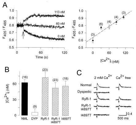
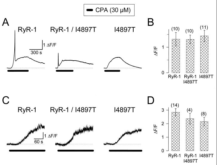
Central core disease (CCD) is a human congenital myopathy characterized by fetal hypotonia and proximal muscle weakness that is linked to mutations in the gene encoding the type-1 ryanodine receptor (RyR1). CCD is thought to arise from Ca(2+)-induced damage stemming from mutant RyR1 proteins forming "leaky" sarcoplasmic reticulum (SR) Ca(2+) release channels. A novel mutation in the C-terminal region of RyR1 (I4898T) accounts for an unusually severe and highly penetrant form of CCD in humans [Lynch, P. J., Tong, J., Lehane, M., Mallet, A., Giblin, L., Heffron, J. J., Vaughan, P., Zafra, G., MacLennan, D. H. & McCarthy, T. V. (1999) Proc. Natl. Acad. Sci. USA 96, 4164--4169]. We expressed in skeletal myotubes derived from RyR1-knockout (dyspedic) mice the analogous mutation engineered into a rabbit RyR1 cDNA (I4897T). Here we show that homozygous expression of I4897T in dyspedic myotubes results in a complete uncoupling of sarcolemmal excitation from voltage-gated SR Ca(2+) release without significantly altering resting cytosolic Ca(2+) levels, SR Ca(2+) content, or RyR1-mediated enhancement of dihydropyridine receptor (DHPR) channel activity. Coexpression of both I4897T and wild-type RyR1 resulted in a 60% reduction in voltage-gated SR Ca(2+) release, again without altering resting cytosolic Ca(2+) levels, SR Ca(2+) content, or DHPR channel activity. These findings indicate that muscle weakness suffered by individuals possessing the I4898T mutation involves a functional uncoupling of sarcolemmal excitation from SR Ca(2+) release, rather than the expression of overactive or leaky SR Ca(2+) release channels.


Figures

Figure 1
Figure 1
The I4897T mutation in RyR1 reduces electrically evoked Ca2+ release without altering resting cytosolic Ca2+ levels. (A) In situ calibration (22) of Indo-1 obtained in intact myotubes. (Left) Three representative experiments in which Indo-1 ratios (F405/F485) were obtained in Indo-1 AM-loaded myotubes before and after (arrow) establishment of the whole cell configuration by using internal solutions containing different levels of free Ca2+. (Right) In situ Indo-1 calibration curve used to determine resting Ca2+ levels. This calibration curve was obtained by using four different BAPTA-buffered calibration solutions (closed triangles). The Indo-1 ratio of myotubes dialyzed with the internal solution (except fluo-3 was replaced with Indo-1) used in patch clamp experiments (Figs. 2 and 3) was superimposed on the fitted curve (open triangle). The free Ca2+ level of this internal solution (≈60 nM) is nearly identical to that of intact normal myotubes (Fig. 1B) and, thus, should be sufficient for maintaining the loading state of the SR. (B) Resting Ca2+ levels determined by using the in situ calibration obtained from normal myotubes (NML), uninjected dyspedic myotubes (DYP), and dyspedic myotubes expressing RyR1 alone (RyR-1), RyR1 and I4897T (RyR-1/I4897T), or I4897T alone (I4897T). (C) Electrically evoked (arrows) Ca2+ transients recorded from Indo-1 AM-loaded myotubes in the presence and absence of extracellular Ca2+.
Figure 2
Figure 2
Homozygous and heterozygous expression of I4897T in dyspedic myotubes does not alter luminal SR Ca2+ levels monitored with Fluo-3. (A) CPA-induced Ca2+ transients in intact, fluo-3 AM-loaded dyspedic myotubes expressing RyR1 alone (Left), RyR1/I4897T (Center), or I4897T alone (Right). (B) Average maximal values of slow CPA-induced Ca2+ transients obtained from intact myotubes (mean ± SEM). Control experiments revealed that 30 μM CPA induced a similar fluorescence increase in intact normal (0.98 ± 0.30, n = 7) and dyspedic (1.07 ± 0.20, n = 7) myotubes. These values were not significantly different from those of RyR1-, I4897T-, or RyR1/I4897T-expressing myotubes (P > 0.3). In addition, RyR1- and I4897T-expressing myotubes exhibited CPA-induced Ca2+ transients of similar magnitude even when elicited in a nominally Ca2+-free external solution. (C) CPA-induced Ca2+ transients measured in whole-cell patch clamped dyspedic myotubes expressing RyR1 alone (Left), RyR1/I4897T (Center), or I4897T alone (Right). (D) Average maximal values of CPA-induced Ca2+ transients obtained from C (mean ± SEM).
Figure 3
Figure 3
The I4897T mutation in RyR1 disrupts voltage-gated SR Ca2+ release but not RyR1-mediated enhancement of DHPR channel activity. (A) Representative L-currents (lower traces) and Ca2+ transients (upper traces) obtained following 30-ms depolarizations to the indicated potentials obtained from uninjected dyspedic myotubes (first column) and dyspedic myotubes expressing RyR1 alone (second column), RyR1/I4897T (third column), or I4897T alone (fourth column). The outward deflection at the beginning of the current traces represents charge movement, which did not differ between uninjected and RyR1-expressing dyspedic myotubes (19, 20). For clarity, the ionic current during the voltage step to +40 mV for the uninjected dyspedic myotube (Inset) and all of the Ca2+ transients for the I4897T-expressing myotube were amplified ten times. Under these recording conditions (0.1 mM EGTA internal solution and 10-s interpulse duration), the baseline fluorescence was similar for all pulses within a sequence, even following pulses that elicit large Ca2+ release. (BD) Average voltage dependence of peak L-current density (B) and Ca2+ transients (C) for dyspedic myotubes expressing RyR1 alone (black circles), RyR1/I4897T (white squares), or I4897T alone (white triangles). (D) Voltage dependence of the Ca2+ transients normalized to their respective peak values. The U-shaped curve through the data for the I4897T-expressing myotubes was obtained by inverting, and normalizing to one, the I–V curve shown in B.

References

    1. MacLennan D H. Eur J Biochem. 2000;267:5291–5297. - PubMed
    1. Berchtold M W, Brinkmeier H, Muntener M. Physiol Rev. 2000;80:1215–1265. - PubMed
    1. Isaacs H, Heffron J J, Badenhorst M. J Neurol Neurosurg Psychiatry. 1975;38:1177–1186. - PMC - PubMed
    1. Shy G M, Magee K R. Brain. 1956;79:610–621. - PubMed
    1. Shuaib A, Paasuke R T, Brownwell K W. J Comp Pathol. 1987;97:597–600. - PubMed

Publication types

Substances