. 2001 Apr;10(4):707-14.
doi: 10.1110/ps.48001.

Catalytic center of an archaeal type 2 ribonuclease H as revealed by X-ray crystallographic and mutational analyses

Affiliations

Catalytic center of an archaeal type 2 ribonuclease H as revealed by X-ray crystallographic and mutational analyses

A Muroya et al. Protein Sci. 2001 Apr.

Abstract

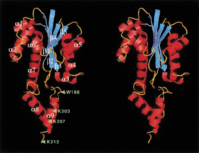
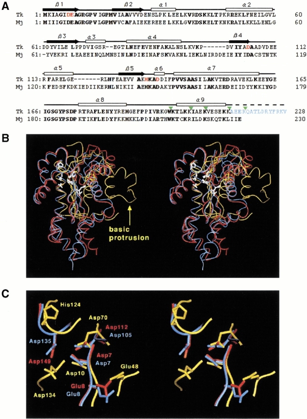
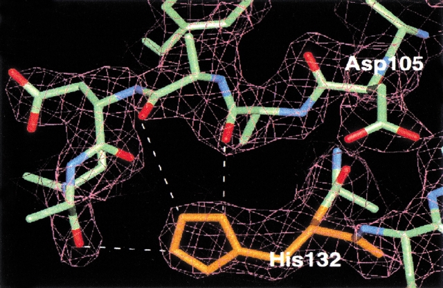
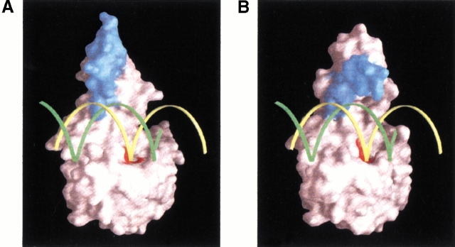
The catalytic center of an archaeal Type 2 RNase H has been identified by a combination of X-ray crystallographic and mutational analyses. The crystal structure of the Type 2 RNase H from Thermococcus kodakaraensis KOD1 has revealed that the N-terminal major domain adopts the RNase H fold, despite the poor sequence similarity to the Type 1 RNase H. Mutational analyses showed that the catalytic reaction requires four acidic residues, which are well conserved in the Type 1 RNase H and the members of the polynucleotidyl transferase family. Thus, the Type 1 and Type 2 RNases H seem to share a common catalytic mechanism, except for the requirement of histidine as a general base in the former enzyme. Combined with the results from deletion mutant analyses, the structure suggests that the C-terminal domain of the Type 2 RNase H is involved in the interaction with the DNA/RNA hybrid.


Figures

Fig. 1.
Fig. 1.
Stereodiagram showing the backbone structure of Tk-RNase HII-213. Helices, strands, and loops are shown in red, cyan, and orange, respectively. Green triangles with labeled amino acids indicate the sites in which the C-terminal truncations (Tk-RNase HII-198, Tk-RNase HII-203, Tk-RNase HII-207, and Tk-RNase HII-213) were constructed.
Fig. 2.
Fig. 2.
(A) Amino acid sequence alignment of Tk-RNase HII (Tk) and Mj-RNase HII (Mj) based on the comparison of their three-dimensional structures. The range of the secondary structures of Tk-RNase HII are shown above the sequence (white box for α-helix and black arrow for β-strand). Bold letters indicate amino acid residues conserved between the two proteins. The amino acid residues shown in red represent the mutated residues in the present study. The amino acid residues shown in blue are the truncated residues 214–228 in Tk-RNase HII-213. The truncation sites are indicated by green triangles. (B) Stereodiagram of the structures of Tk-RNase HII-213 (blue), Mj-RNase HII (red), and Ec-RNase HI (yellow). Root mean square displacements of the superimposition on the Tk-RNase HII structure are 1.2 Å for the 190 Cα atoms of Mj-RNase HII, and 2.0 Å for the 47 Cα atoms consisting of the core β-sheet of Ec-RNase HI. Active-site residues are shown by white stick models. The "basic protrusion" of Ec-RNase HI is indicated by a yellow arrow. (C) A close-up view of the active-site of the three RNases H. The view direction is the same as in the panel B and in Figure 1 ▶.
Fig. 3.
Fig. 3.
A representative 2Fo-Fc electron density map after refinement. The model of a protein portion around His132 is fitted into density contoured at the 1.0 σ level. The imidazole group can assume two conformations, both of which form hydrogen bond(s) with the loop adjacent to the catalytic residue Asp105.
Fig. 4.
Fig. 4.
Surface plasmon resonance analysis of the proteins (100 nM) as reported by Haruki and coworkers (1997). Black, blue, green, orange, red, and cyan lines are sensorgrams for the wild-type protein, Tk-RNase HII-217, Tk-RNase HII-213, Tk-RNase HII-207, Tk-RNase HII-203, and Tk-RNase HII-198, respectively.
Fig. 5.
Fig. 5.
(A) A hypothetical docking model of the DNA/RNA hybrid on Tk-RNase HII-213. This model was generated by superposition of the obtained crystal structure onto the Ec-RNase HI-DNA/RNA model proposed by Nakamura and coworkers (1991). Yellow and green ribbons indicate RNA and DNA strands, respectively. Blue and red surfaces represent the segment 199–213 and the active-site residues, respectively. (B) A putative substrate recognition by Tk-RNase HII. The C-terminal domain is rotated by ∼70° so that the basic α9 helical segment can make contact with phosphate backbones of the DNA/RNA hybrid duplex.

References

    1. Ariyoshi, M., Vassylyev, D.G., Iwasaki, H., Nakamura, H., Shinagawa, H., and Morikawa, K. 1994. Atomic structure of the RuvC resolvase: A Holliday junction-specific endonuclease from E. coli. Cell 82 209–220. - PubMed
    1. Brünger, A.T., Adams, P.D., Clore, G.M., DeLano, W.L., Gros, P., Grosse-Kunstleve, R.W., Jiang, J.S., Kuszewski, J., Nilges, M., Pannu, N.S., et al. 1998. Crystallography and NMR system (CNS): A new software suite for macromolecular structure determination. Acta Cryst. D54 905–921. - PubMed
    1. Bujacz, G., Jaskolski, M., Alexandratos, J., and Wlodawer, A. 1995. High-resolution structure of the catalytic domain of avian sarcoma virus integrase. J. Mol. Biol. 253 333–346. - PubMed
    1. Collaborative Computational Project, Number 4. 1994. The CCP4 suite: Programs for protein crystallography. Acta Cryst. D50 760–763. - PubMed
    1. Cowtan, K.D. 1994. "dm": An automated procedure for phase improvement by density modification. Joint CCP4 and ESF-EACBM Newsl. Protein Crystallogr. 31 34– 38.

MeSH terms

LinkOut - more resources