. 2001 May 25;276(21):17994-9.
doi: 10.1074/jbc.M009327200. Epub 2001 Feb 22.

An essential role of Glu-243 and His-239 in the phosphotransfer reaction catalyzed by pyruvate dehydrogenase kinase

Affiliations

An essential role of Glu-243 and His-239 in the phosphotransfer reaction catalyzed by pyruvate dehydrogenase kinase

A Tuganova et al. J Biol Chem. .

Abstract

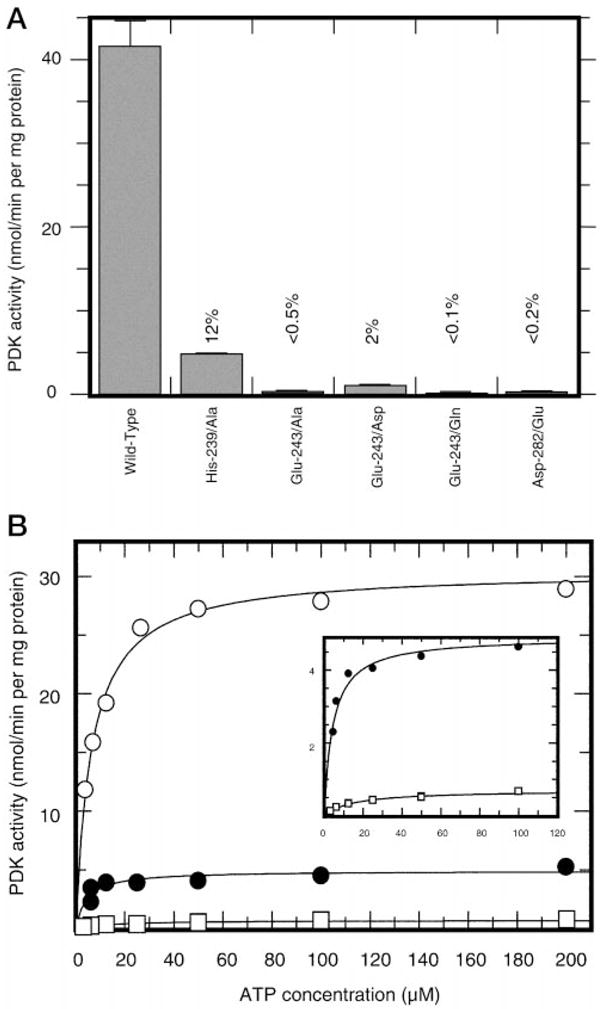
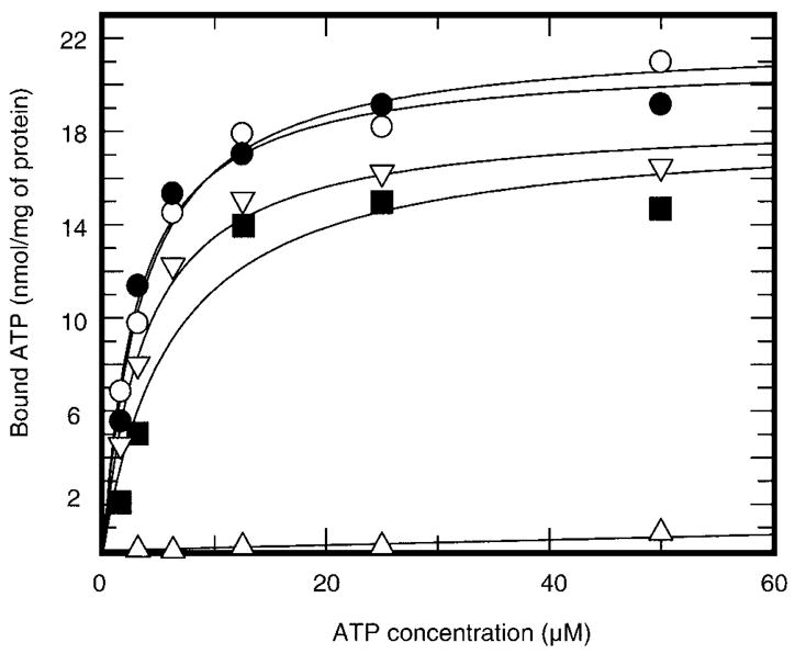
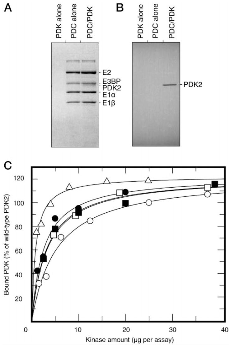
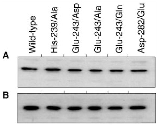
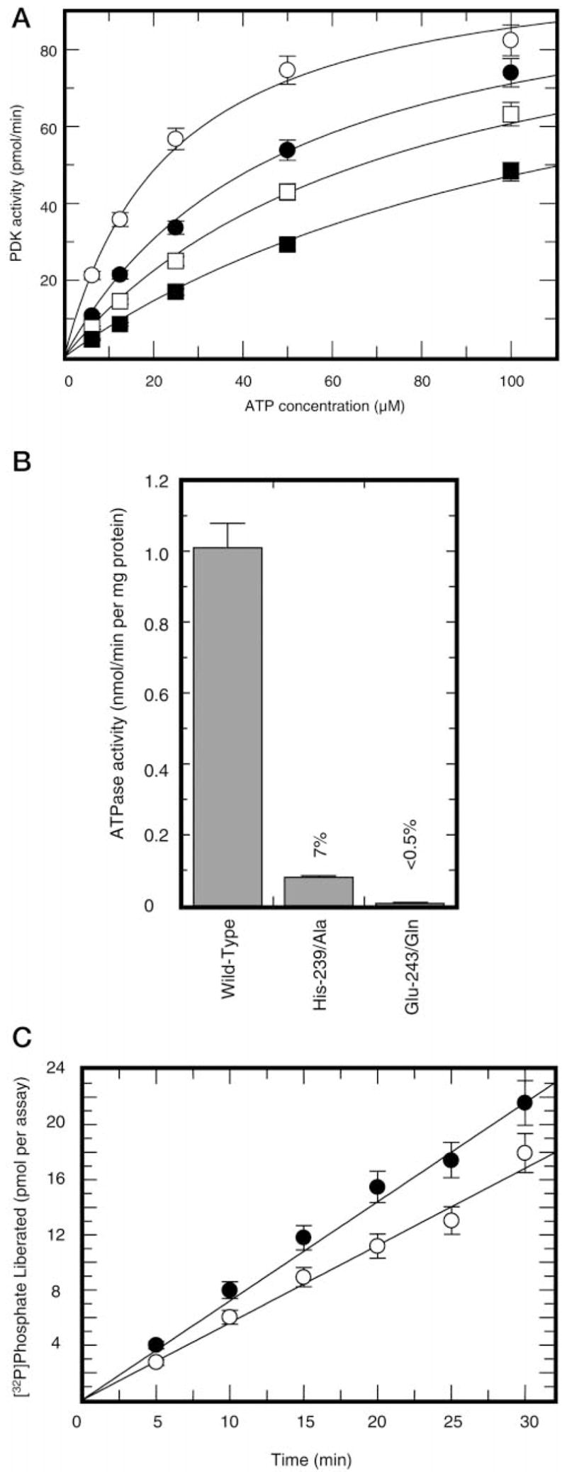
This study was undertaken to examine the mechanistic significance of two highly conserved residues positioned in the active site of pyruvate dehydrogenase kinase, Glu-243 and His-239. We used site-directed mutagenesis to convert Glu-243 to Ala, Asp, or Gln and His-239 to Ala. The resulting mutant kinases demonstrated a greatly reduced capacity for phosphorylation of pyruvate dehydrogenase. The Glu-243 to Asp mutant had approximately 2% residual activity, whereas the Glu-243 to Ala or Gln mutants exhibited less than 0.5 and 0.1% residual activity, respectively. Activity of the His-239 to Ala mutant was decreased by approximately 90%. Active-site titration with [alpha-(32)P]ATP revealed that neither Glu-243 nor His-239 mutations affected nucleotide binding. All mutant kinases showed similar or even somewhat greater affinity than the wild-type kinase toward the protein substrate, pyruvate dehydrogenase complex. Furthermore, neither of the mutations affected the inter-subunit interactions. Finally, pyruvate dehydrogenase kinase was found to possess a weak ATP hydrolytic activity, which required Glu-243 and His-239 similar to the kinase activity. Based on these observations, we propose a mechanism according to which the invariant glutamate residue (Glu-243) acts as a general base catalyst, which activates the hydroxyl group on a serine residue of the protein substrate for direct attack on the gamma phosphate. The glutamate residue in turn might be further polarized through interaction with the neighboring histidine residue (His-239).


Figures

Fig. 1
Fig. 1. Kinetics of PDC phosphorylation by wild-type and mutant PDKs
Panel A, average activities of various PDK preparations analyzed in this study. Experiments were conducted with 200 μM ATP and 1.0 mg/ml recombinant PDC as described under “Experimental Procedures” section. Respective enzymes, except for the wild-type PDK2, were used at final concentrations of 20 μg/ml. The activity of wild-type kinase was calculated based on incorporation of [32P]phosphate during the first 30 s of the reaction. Activities of mutant kinases were calculated based on the incorporation of [32P]phosphate during 30 min of the reaction. Panel B, representative results showing ATP dependence of PDC phosphorylation by wild-type PDK2 (○) and His-239 → Ala (●) and Glu-243 → Asp (□) mutants. The insert shows respective curves for His-239 → Ala (●) and Glu-243 → Asp (□) mutants. ATP concentration was varied from 3 to 200 μM.
Fig. 2
Fig. 2. [α-32P]ATP binding by the wild-type PDK2 and mutant enzymes
Recombinant PDK2, wild-type (○), His-239 → Ala (●) Glu-243 → Ala (▽), Glu-243 → Gln (■), or Asp-282 → Glu (Δ) mutants were incubated with the indicated concentrations of [α-32P]ATP (specific radioactivity ~200 cpm/pmol) for 2 min. Free and protein-bound nucleotides were separated using a vacuum-filtration assay as described under “Experimental Procedures.” The specific binding was determined after subtraction of nonspecific binding from the total binding. The nonspecific binding was determined in the presence of 1,000-fold excess of cold ATP.
Fig. 3
Fig. 3. Binding of wild-type PDK2 and respective mutant kinases to recombinant PDC
Panel A, SDS/PAGE analysis of wild-type PDK2, recombinant PDC, and PDC-kinase complex centrifuged through Sephacel S300 columns. Panel B, Western blot analysis of corresponding preparations with monoclonal antibodies against His6-tag. Immunoreactive bands were visualized by [125I]protein A staining followed by autoradiography. Panel C, concentration-dependence curves for binding of wild-type PDK2 (○), His-239 → Ala (□), Glu-239 → Asp (■), Glu-243 → Gln (●), and Asp-282 → Glu (△) mutants to recombinant PDC. Binding curves were constructed based on the results of scanning densitometry of the respective Western blots stained with 125I-protein A. Data was analyzed using UN-SCAN-IT gel™ software (Silk Scientific, Inc., Orem, UT). Results are expressed as percent of wild-type PDK2 bound to PDC.
Fig. 4
Fig. 4. Western blot analysis of differentially tagged preparations of PDK purified by metal affinity chromatography
Panel A, affinity-purified kinase preparations probed with antibodies raised against T7-tag. Panel B, corresponding preparations probed with anti-H6-tag antibodies. Immunoreactive bands were visualized by 125I-protein A staining followed by autoradiography.
Fig. 5
Fig. 5. ATP hydrolytic activity in preparations of PDK2
Panel A, inhibition of pyruvate dehydrogenase kinase activity by radicicol. Kinase activity was determined without radicicol (○) or in the presence of radicicol at concentrations of 25 μM (●), 50 μM (■), or 100 μM (□). Panel B, radicicol-sensitive ATP hydrolytic activity in preparations of wild-type, Glu-243 → Gln, and His-239 → Ala PDK2. Total ATP hydrolysis was measured in the absence of radicicol with 10 μM [γ-32P]ATP as a substrate. Nonspecific hydrolysis was determined in the presence of 200 μM radicicol. Shown is radicicol-sensitive ATPase activity (difference between total and nonspecific activity) intrinsic to PDK2. Panel C, radicicol-sensitive (○) versus E1-sensitive (●) ATPase activity in preparations of wild-type PDK2. Radicicol-sensitive ATPase activity was determined essentially as described in the legend to panel B. E1-sensitive ATPase activity was determined as a difference between total ATPase activity and ATPase activity measured in the presence of E1 component (final concentration 1.0 mg/ml).

References

    1. Randle PJ. Proc Nutr Soc. 1995;54:317–327. - PubMed
    1. Linn TC, Pettit FH, Reed LJ. Biochemistry. 1968;62:234–241.
    1. Stepp LR, Pettit FH, Yeaman SJ, Reed LJ. J Biol Chem. 1983;258:9454–9458. - PubMed
    1. Teague WM, Pettit FH, Wu TL, Silberman SR, Reed LJ. Biochemistry. 1982;21:5585–5592. - PubMed
    1. Popov KM, Kedishvili NY, Zhao Y, Gudi R, Harris RA. J Biol Chem. 1994;269:29720–29724. - PubMed

Publication types

LinkOut - more resources