. 2001 May 18;276(20):17515-23.
doi: 10.1074/jbc.M009731200. Epub 2001 Feb 8.

The missing link in coronavirus assembly. Retention of the avian coronavirus infectious bronchitis virus envelope protein in the pre-Golgi compartments and physical interaction between the envelope and membrane proteins

Affiliations

The missing link in coronavirus assembly. Retention of the avian coronavirus infectious bronchitis virus envelope protein in the pre-Golgi compartments and physical interaction between the envelope and membrane proteins

K P Lim et al. J Biol Chem. .

Abstract

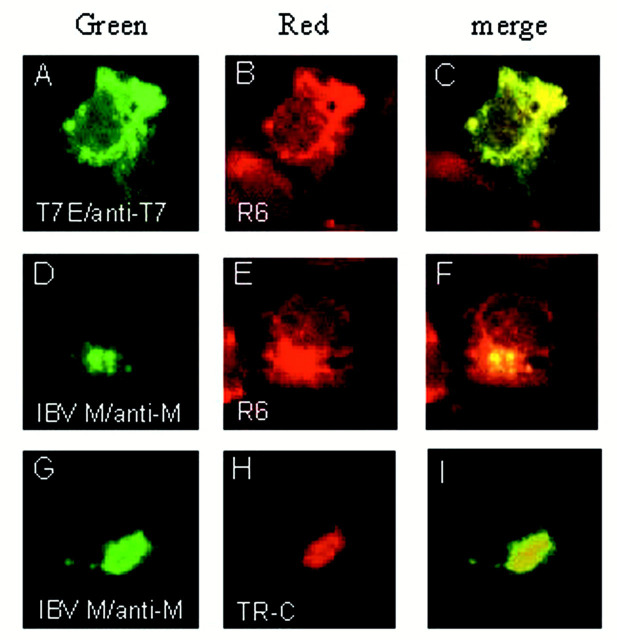
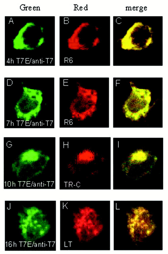
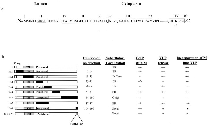
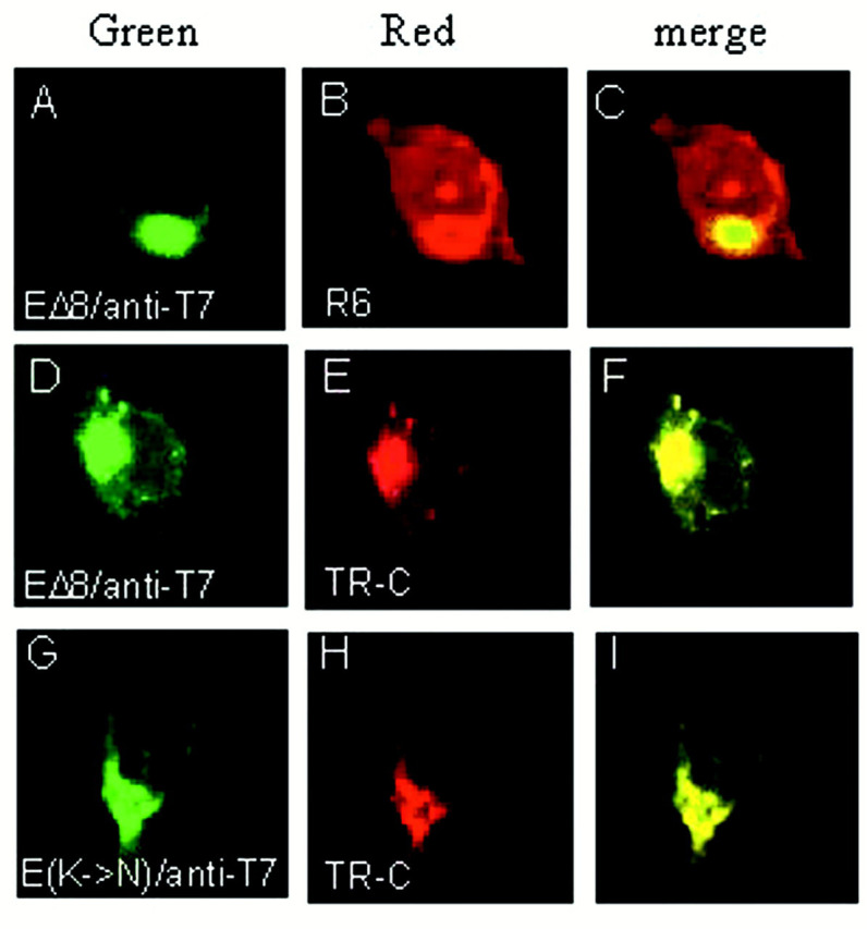
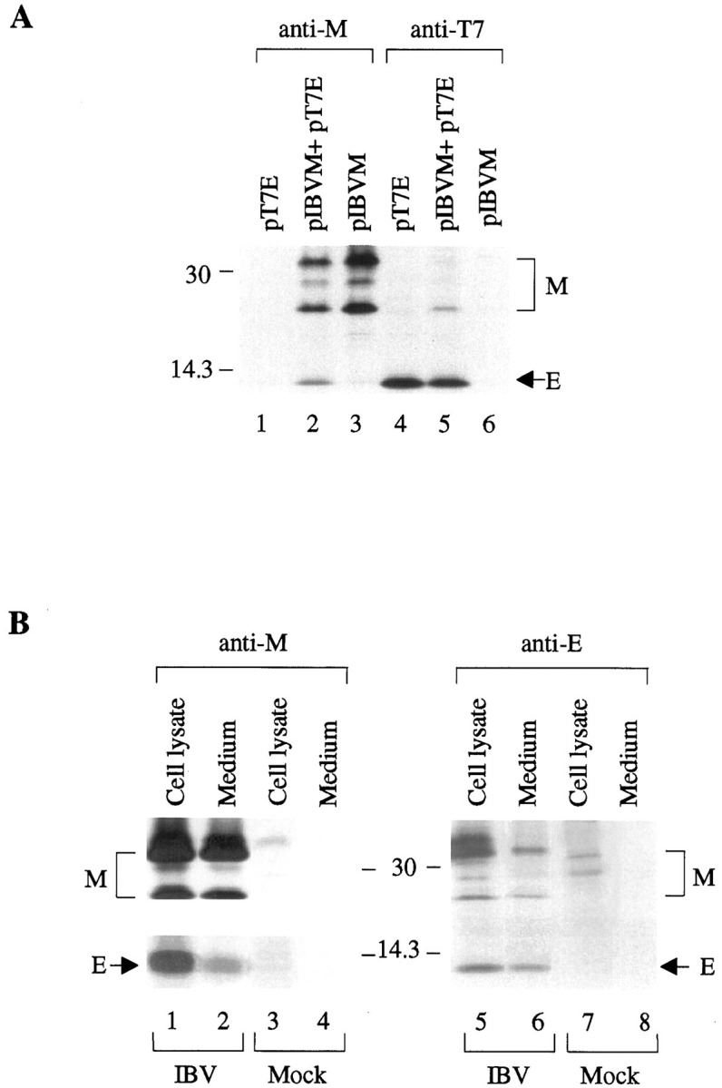
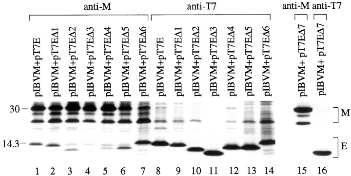
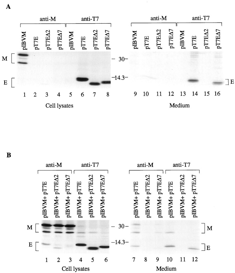
One missing link in the coronavirus assembly is the physical interaction between two crucial structural proteins, the membrane (M) and envelope (E) proteins. In this study, we demonstrate that the coronavirus infectious bronchitis virus E can physically interact, via a putative peripheral domain, with M. Deletion of this domain resulted in a drastic reduction in the incorporation of M into virus-like particles. Immunofluorescent staining of cells coexpressing M and E supports that E interacts with M and relocates M to the same subcellular compartments that E resides in. E was retained in the pre-Golgi membranes, prior to being translocated to the Golgi apparatus and the secretory vesicles; M was observed to exhibit similar localization and translocation profiles as E when coexpressed with E. Deletion studies identified the C-terminal 6-residue RDKLYS as the endoplasmic reticulum retention signal of E, and site-directed mutagenesis of the -4 lysine residue to glutamine resulted in the accumulation of E in the Golgi apparatus. The third domain of E that plays a crucial role in virus budding is a putative transmembrane domain present at the N-terminal region, because deletion of the domain resulted in a free distribution of the mutant protein and in dysfunctional viral assembly.


Figures

Figure 1
Figure 1
Subcellular localization of IBV E and M proteins in transfected Cos-7 cells. The T7-tagged E and M expressed in Cos-7 cells were detected using monoclonal anti-T7 (A) and polyclonal anti-M antibodies (D andG), respectively. The proteins were then labeled with the FITC-conjugated secondary antibodies. Immunofluorescent staining of E (A) and M (D and G) gives a reticular staining pattern and a perinuclear, Golgi-like staining pattern, respectively. B and E refer to cells stained with R6, a dye for the ER, and H refers to a cell stained with BODIPY TR-ceramide (TR-C), a vital dye for the Golgi apparatus. The green images represent the FITC-derived green fluorescence, and red images represent the rhodamine and Texas Red-derived red fluorescence. Colocalization of viral proteins with the organelle markers is represented by theyellow region within each cell in the merged images (C, F, and I). The fluorescence was viewed using a confocal scanning Zeiss microscope.
Figure 2
Figure 2
Subcellular localization and translocation of E protein. Cos-7 cells expressing E were incubated in the presence of 100 μg/ml cycloheximide for 30 min at 3.5 h post-transfection. The cells were fixed at 4, 7, 10, and 16 h and subjected to indirect immunofluorescent staining with anti-T7 monoclonal antibody followed by incubating with FITC-conjugated anti-mouse antiserum. Immunostaining of E is presented in A,D, G, and J. B andE show the ER staining with R6; H shows the BODIPY TR-ceramide (TR-C) staining pattern; and Kshows the LysoTrackerTM Red DND-99 (LT) staining pattern. The green images represent the FITC-derived green fluorescence, and red images represent the Rhodamine and Texas Red-derived red fluorescence. Colocalization of viral proteins with the organelle markers is represented by theyellow region within each cell in the merged images (C, F, I, and L).
Figure 3
Figure 3
a, diagram indicating the presence of four putative domains in IBV E. E adopts a type III topology. Theboxed regions refer to the putative domains. Irefers to a potential N-linked glycosylation site which is not utilized (15), II refers to a putative transmembrane domain between residues 17 and 33, III refers to a putative peripheral domain located between amino acids 37 and 53, and IV refers to a potential ER retention signal. b, summary of the effects of deletion and mutation on the subcellular localization of IBV E, the interaction with M, VLP release, and the incorporation of M into VLPs. The 109 amino acids of E are represented by white boxes, and the putative transmembrane (TM) and peripheral domains are indicated in gray boxes. The deleted regions are represented in black boxes, and a single mutation is represented by a stippled box. RDKLYSstands for the cytoplasmic tail sequence of E containing the potential ER retention signal. Also included is a summary of the position of deletion, subcellular localization of each mutant, coimmunoprecipitation with M, VLP release, and the incorporation of M into the wild type and mutant E-induced VLPs.
Figure 4
Figure 4
The effects of deletions on the subcellular localization of E protein. Cos-7 cells expressing the wild type and mutant E were fixed at 7 h post-transfection. The expressed proteins were detected with anti-T7 monoclonal antibody against the T7 tag fused to the N terminus of the wild type E (A), EΔ1 (D), EΔ2 (G), EΔ3 (J), EΔ4 (M), EΔ5 (P), and EΔ6 (S).B, E, H, K, N,Q, and T refer to cells stained with R6. Thegreen images represent the FITC-derived green fluorescence, and red images represent the Rhodamine and Texas Red-derived red fluorescence. The merged images (C, F, I, L, O, R, andU; yellow) represent colocalization of proteins with the ER marker.
Figure 5
Figure 5
Subcellular localization of E proteins lacking the putative ER retention signal. Cos-7 cells transiently expressing EΔ8 (A and D) and E(K→N) (G) were fixed at 7 h post-transfection. Proteins are detected with anti-T7 monoclonal antibody. B refers to cells stained with R6, and E and H show cells stained with BODIPY TR-ceramide (TR-C). The green images represent the FITC-derived green fluorescence, and redimages represent the Rhodamine and Texas Red-derived red fluorescence. The merged images (yellow) represent colocalization of proteins with the organelle markers (C, F, andI).
Figure 6
Figure 6
a, coimmunoprecipitation of M and E proteins in Cos-7 cells expressing the two proteins. Cos-7 cells expressing E (lanes 1 and 4), M (lanes 3 and 6), or coexpressing both proteins (lanes 2 and 5) were lysed and subjected to immunoprecipitation before analysis on SDS-17.5% polyacrylamide gel. The expressed proteins were immunoprecipitated with anti-M (lanes 1–3) and anti-T7 (lanes 4–6) antisera, respectively.Numbers on the left indicate molecular masses in kilodaltons. b, coimmunoprecipitation of M and E proteins in IBV-infected Vero cells. Cell lysates were prepared from Vero cells harvested at 18 h post-infection, and the collected culture media were precleared by centrifugation. The viral proteins in the cell lysates (lanes 1, 3, 5, and7) and in the culture medium (lanes 2,4, 6, and 8) were immunoprecipitated with anti-M (lanes 1–4) and anti-E (lanes 5–8), before analysis on SDS-17.5% polyacrylamide gel. The upper part of the left panel was prepared from a gel exposed for 1 day, and the lower part was prepared from the same gel exposed for 3 days. Numbers on the left indicate molecular masses in kilodaltons.
Figure 7
Figure 7
The effects of coexpression of E with M on the subcellular localization of M. M was coexpressed with E in Cos-7 cells, incubated in the presence of 100 μg/ml of cycloheximide for 30 min, and subjected to indirect immunofluorescent staining. The cells were fixed at 4 (A–C), 7 (D–F), 10 (G–I), and 16 (J–L) h, respectively, and the subcellular localization of proteins were examined by dual labeling with a mixture of anti-T7 (mouse) and anti-M (rabbit) antisera, followed by incubating with a mixture of FITC-conjugated anti-mouse and tetramethyl rhodamine isocyanate-conjugated anti-rabbit antisera. The staining patterns of E are indicated by the green images (A, D, G, and J), and the staining patterns of M are indicated by the red images (B, E, H, K, N, and Q). M and P refer to cells expressing EΔ2 and EΔ7, respectively. Merged images (yellow) represent colocalization of the two proteins (C, F, I, L, O, and R).
Figure 8
Figure 8
Subcellular localization of IBV E and M proteins in IBV infected cells. Vero cells infected with IBV were incubated in the presence of 100 μg/ml cycloheximide for 30 min at 4.5 h post-infection and subjected to indirect immunofluorescent staining. The cells were fixed at 5 (A–C andM–O), 7 (D–F and P–R), 9 (G–I and S–U), and 12 (J–L andV–X) h post-infection, respectively. Viral proteins were detected with anti-E (A, D, G, andJ) and anti-M (M, P, S, andV) antisera. B, E, N, andQ refer to ER staining with R6; H andT show cells stained with BODIPY TR-ceramide (TR-C); and K and W show cells stained with LysoTrackerTM Red DND-99 (LT). Thegreen images represent the FITC-derived green fluorescence, and red images represent the rhodamine and Texas Red-derived red fluorescence. The merged images (yellow) represent colocalization of the proteins with the organelle markers (C, F, I, L, O,R, U, and X).
Figure 9
Figure 9
Coimmunoprecipitation of M protein and E deletion mutants from transfected Cos-7 cells. Cos-7 cells were transfected with plasmids expressing M and the wild type and mutant E, as indicated above each lane. The cells were lysed and subjected to immunoprecipitation before analysis with SDS-17.5% polyacrylamide gel.Lanes 1–7 and 15 refer to products precipitated with anti-M, and lanes 8–14 and 16 refer to products precipitated with anti-T7. Numbers on theleft indicate molecular masses in kilodaltons.
Figure 10
Figure 10
a, the effects of deletion of the putative transmembrane and the peripheral membrane domains of E on the release of VLPs. Cos-7 cells were transfected with plasmids as indicated above each lane. Cell lysates were prepared and subjected to immunoprecipitation with anti-M (lanes 1–4) and anti-T7 (lanes 5–8), and protein expression efficiency was analyzed on SDS-17.5% polyacrylamide gel. The release of VLP was analyzed by immunoprecipitation of the culture medium from each transfection with anti-M (lanes 9–12) and anti-T7 (lanes 13–16) after the medium was centrifuged at low speed (4,000 ×g) to preclear the cell debris. The numbersbetween the two panels indicate molecular masses in kilodaltons.b, the effects of deletion of the putative transmembrane and the peripheral membrane domains of E on the incorporation of M into VLPs. Cos-7 cells were transfected with plasmids as indicated above each lane. Cell lysates were prepared and subjected to immunoprecipitation with anti-M (lanes 1–3) and anti-T7 (lanes 4–6), and protein expression efficiency was analyzed on SDS-17.5% polyacrylamide gel. The release of VLP was analyzed by immunoprecipitation of the culture medium from each transfection with anti-M (lanes 7–9) and anti-T7 (lanes 10–12) after the medium was centrifuged at low speed (4,000 ×g) to preclear the cell debris. The numbersbetween the two panels indicate molecular masses in kilodaltons.

Similar articles

Cited by

References

    1. Brautigam S., Snezhkov E., Bishop D.H. Virology. 1993;192:512–524. - PubMed
    1. Vennema H., Godeke G.-J., Rossen J.W.A., Voorhout W.F., Horzinek M.C., Opstelten D.-J., E., Rottier P.J.M. EMBO J. 1996;15:2020–2028. - PMC - PubMed
    1. Li T.C., Yamakawa Y., Suzuki K., Tatsumi M., Razak M.A., Uchida T., Takeda N., Miyamura T. J. Virol. 1997;71:7207–7213. - PMC - PubMed
    1. White L.J., Hardy M.E., Estes M.K. J. Virol. 1997;71:8066–8072. - PMC - PubMed
    1. Baumert T.F., Ito S., Wong D.T., Liang T.J. J. Virol. 1998;72:3827–3836. - PMC - PubMed

Publication types

MeSH terms

LinkOut - more resources