

Phenotypic and genotypic characterization of Pediococcus strains isolated from human clinical sources
- PMID: 11283035
- PMCID: PMC87918
- DOI: 10.1128/JCM.39.4.1241-1246.2001
Phenotypic and genotypic characterization of Pediococcus strains isolated from human clinical sources
Abstract
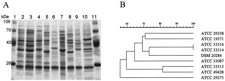
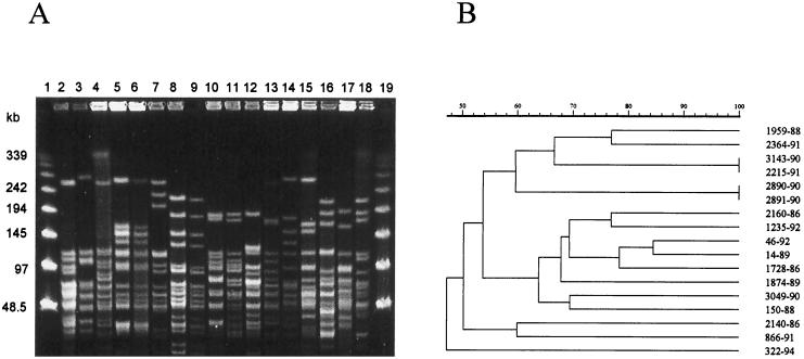
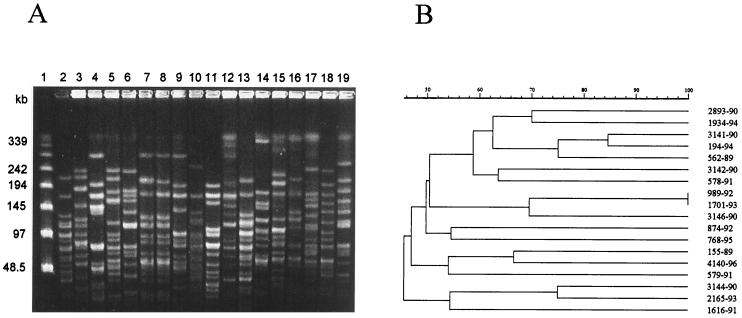
Seventy-two strains of pediococci isolated from human clinical sources were characterized by conventional physiological tests, chromogenic enzymatic tests, analysis of whole-cell protein profiles (WCPP) by sodium dodecyl sulfate-polyacrylamide gel electrophoresis, and analysis of chromosomal DNA restriction profiles by pulsed-field gel electrophoresis (PFGE). Conventional tests allowed identification of 67 isolates: 52 strains were identified as Pediococcus acidilactici, 15 strains were identified as Pediococcus pentosaceus, and 5 strains were not identified because of atypical reactions. Analysis of WCPP identified all isolates since each species had a unique WCPP. By the WCPP method, the atypical strains were identified as P. acidilactici (two strains) and P. pentosaceus (three strains). The chromogenic substrate test with o-nitrophenyl-beta-D-glucopyranoside differentiated all 54 strains of P. acidilactici (negative reactions) and 13 (72%) of 18 strains of P. pentosaceus (positive reactions). Isolates of both species were shown to be nonclonal as revealed by the genetic diversity when chromosomal DNA was analyzed by PFGE. Using WCPP as the definitive identification procedure, P. acidilactici (28 of 54 strains; 51.8%) was more likely than P. pentosaceus (4 of 18 strains; 22.3%) to be isolated from blood cultures.
Figures
Similar articles
-
Pediococcus stilesii sp. nov., isolated from maize grains.Int J Syst Evol Microbiol. 2006 Feb;56(Pt 2):329-333. doi: 10.1099/ijs.0.63944-0. Int J Syst Evol Microbiol. 2006. PMID: 16449434
-
Phenotypic and genotypic characterization of Thai oral streptococci, lactobacilli and pediococci.Trop Biomed. 2012 Jun;29(2):254-64. Trop Biomed. 2012. PMID: 22735847
-
Pediococcus lolii sp. nov., isolated from ryegrass silage.Int J Syst Evol Microbiol. 2009 May;59(Pt 5):1007-10. doi: 10.1099/ijs.0.005793-0. Int J Syst Evol Microbiol. 2009. PMID: 19406783
-
Autolytic activity and pediocin-induced lysis in Pediococcus acidilactici and Pediococcus pentosaceus strains.J Appl Microbiol. 2003;94(4):561-70. doi: 10.1046/j.1365-2672.2003.01868.x. J Appl Microbiol. 2003. PMID: 12631191
-
Genotypic and phenotypic diversity of Pediococcus pentosaceus strains isolated from food matrices and characterisation of the penocin operon.Antonie Van Leeuwenhoek. 2013 May;103(5):1149-63. doi: 10.1007/s10482-013-9897-1. Epub 2013 Feb 27. Antonie Van Leeuwenhoek. 2013. PMID: 23444039
Cited by
-
Effect of sodium selenite on the bacteria growth, selenium accumulation, and selenium biotransformation in Pediococcus acidilactici.Food Sci Biotechnol. 2017 Jul 20;26(4):1013-1018. doi: 10.1007/s10068-017-0142-y. eCollection 2017. Food Sci Biotechnol. 2017. PMID: 30263631 Free PMC article.
-
Genomic diversity within the genus Pediococcus as revealed by randomly amplified polymorphic DNA PCR and pulsed-field gel electrophoresis.Appl Environ Microbiol. 2002 Feb;68(2):765-71. doi: 10.1128/AEM.68.2.765-771.2002. Appl Environ Microbiol. 2002. PMID: 11823217 Free PMC article.
-
Molecular identification of lactic acid bacteria SR6 strain and evaluation of its activity as an anticancer in T47D cell line.Vet World. 2022 Jun;15(6):1583-1588. doi: 10.14202/vetworld.2022.1583-1588. Epub 2022 Jun 29. Vet World. 2022. PMID: 35993063 Free PMC article.
-
Miscellaneous catalase-negative, gram-positive cocci: emerging opportunists.J Clin Microbiol. 2002 Apr;40(4):1129-33. doi: 10.1128/JCM.40.4.1129-1133.2002. J Clin Microbiol. 2002. PMID: 11923320 Free PMC article. Review. No abstract available.
-
Mucosa-adherent Pediococcus Pentosaceus I44 isolated from healthy human and effect of oleic acid on its probiotic properties.Curr Res Microb Sci. 2021 Aug 14;2:100058. doi: 10.1016/j.crmicr.2021.100058. eCollection 2021 Dec. Curr Res Microb Sci. 2021. PMID: 34841348 Free PMC article.
References
-
- Back W, Stackebrandt E. DNS/DNS homologiestudien innerhalb der Gattung Pediococcus. Arch Microbiol. 1978;118:79–85.
-
- Collins M D, Williams A M, Wallbanks S. The phylogeny of Aerococcus and Pediococcus as determined by 16S rRNA sequences analysis: description of Tetragenococcus gen. nov. FEMS Microbiol Lett. 1990;70:255–262. - PubMed
-
- Colman G, Efstratiou A. Vancomycin-resistant leuconostoc, lactobacilli and now pediococci. J Hosp Infect. 1987;10:1–3. - PubMed
-
- Corcoran G D, Gibbons N, Mulvihill T E. Septicaemia caused by Pediococcus pentosaceus: a new opportunistic pathogen. J Infect. 1991;23:179–182. - PubMed
MeSH terms
Substances
LinkOut - more resources
Full Text Sources
Other Literature Sources
Molecular Biology Databases
