. 2001 Apr;39(4):1353-9.
doi: 10.1128/JCM.39.4.1353-1359.2001.

Molecular characterization of invasive and noninvasive Campylobacter jejuni and Campylobacter coli isolates

Affiliations

Molecular characterization of invasive and noninvasive Campylobacter jejuni and Campylobacter coli isolates

A C Carvalho et al. J Clin Microbiol. 2001 Apr.

Abstract

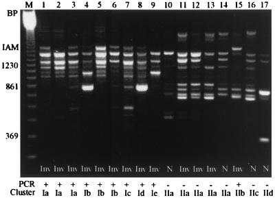
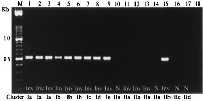
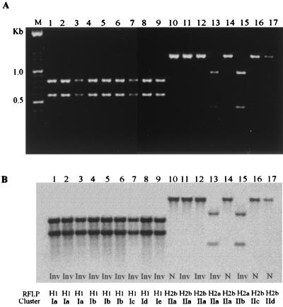
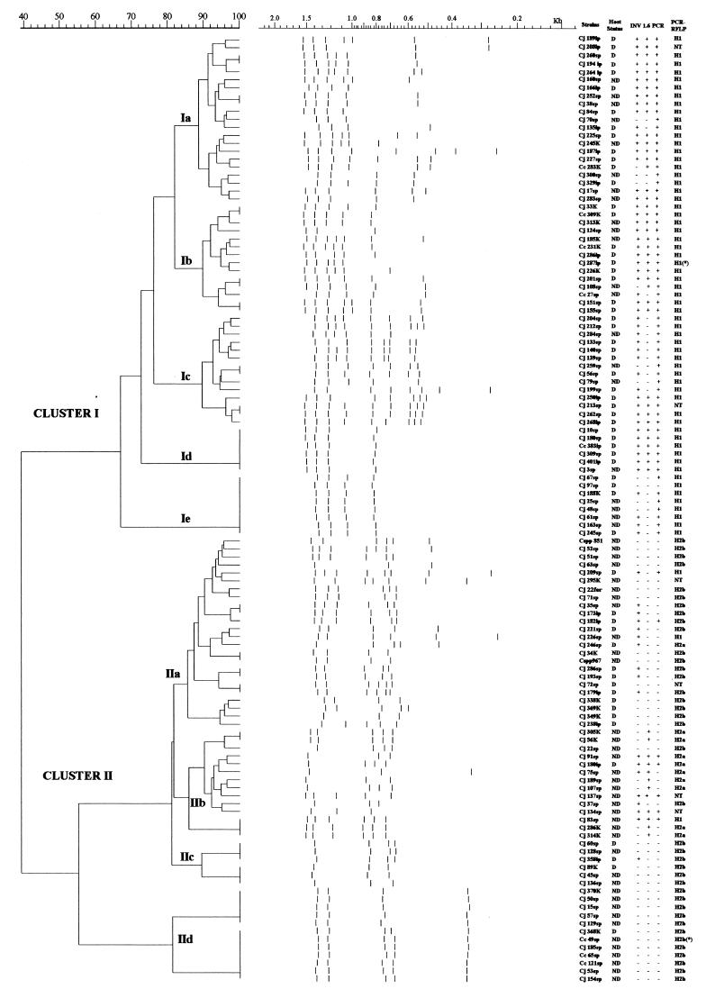
Campylobacter jejuni is one of the most common causes of bacterial diarrhea worldwide and is the primary bacterial cause of food-borne illness. Adherence to and invasion of epithelial cells are the most important pathogenic mechanisms of Campylobacter diarrhea. Molecular characterization of invasive and noninvasive Campylobacter isolates from children with diarrhea and symptom-free children was performed by random amplified polymorphic DNA techniques (RAPD). A distinct RAPD profile with a DNA band of 1.6 kb was observed significantly more frequently among invasive (63%) than among noninvasive (16%) Campylobacter isolates (P = 0.000005). The 1.6-kb band was named the invasion-associated marker (IAM). Using specifically designed primers, a fragment of 518 bp of the iam locus was amplified in 85% of invasive and 20% of noninvasive strains (P = 0.0000000). Molecular typing with a PCR-restriction fragment length polymorphism assay which amplified the entire iam locus showed a HindIII restriction fragment polymorphism pattern associated mainly with invasive strains. Although cluster analysis of the RAPD fingerprinting showed genetic diversity among strains, two main clusters were identified. Cluster I comprised significantly more pathogenic and invasive isolates, while cluster II grouped the majority of nonpathogenic, noninvasive isolates. These data indicate that most of the invasive Campylobacter strains could be differentiated from noninvasive isolates by RAPD analysis and PCR using specific primers that amplify a fragment of the iam locus.


Figures

FIG. 1
FIG. 1
RAPD amplification products of invasive and noninvasive Campylobacter strains using the arbitrary primer 1290. The IAM band size was estimated as 1.6 kb, marked on the left side. Results of the iam PCR amplification and their location in cluster analysis is shown. Lanes: M, 123-bp ladder marker; 1, C. jejuni 84sp; 2, C. jejuni 135ip; 3, C. jejuni 227sp; 4, C. jejuni 33K; 5, C. jejuni 287ip; 6, C. jejuni 151sp; 7, C. jejuni 268ip; 8, C. jejuni 401ip; 9, C. jejuni 188K; 10, C. jejuni 63sp; 11, C. jejuni 221sp; 12, C. jejuni 286sp; 13, C. jejuni 246sp; 14, C. jejuni 349K; 15, C. jejuni 180ip; 16, C. jejuni 128sp; 17, C. coli 49sp. Inv, invasive strains; N, noninvasive strains.
FIG. 2
FIG. 2
iam PCR amplification, using primers 1.6F and 1.6R, of the genomic DNA of noninvasive and invasive Campylobacter strains. The location in cluster analysis is shown. Lanes: M, 100-bp ladder; 1, C. jejuni 84sp; 2, C. jejuni 135ip; 3, C. jejuni 227sp; 4, C. jejuni 33K; 5, C. jejuni 287ip; 6, C. jejuni 151sp; 7, C. jejuni 268ip; 8, C. jejuni 401ip; 9, C. jejuni 188K; 10, C. jejuni 63sp; 11, C. jejuni 221sp; 12, C. jejuni 286sp; 13, C. jejuni 246sp; 14, C. jejuni 349K; 15, C. jejuni 180ip; 16, C. jejuni 128sp; 17, C. coli 49sp; 18, negative control.
FIG. 3
FIG. 3
(A) Electrophoresis showing PCR-RFLP analysis of the iam locus. An ∼1,360-bp PCR product amplified with degenerated primers (p77F and p1415R) was digested with HindIII, and three patterns were identified: H1 (lanes 1 to 9), H2a (lanes 13 to 15), and H2b (lanes 10 to 12, 14, 16, and 17). (B) Southern blot of the same gel with the entire 1.6-kb probe labeled with DIG. Lanes: M, 100-bp ladder; 1, C. jejuni 84sp; 2, C. jejuni 135ip; 3, C. jejuni 227sp; 4, C. jejuni 33K; 5, C. jejuni 287ip; 6, C. jejuni 151sp; 7, C. jejuni 286ip; 8, C. jejuni 401ip; 9, C. jejuni 188K; 10, C. jejuni 63sp; 11, C. jejuni 221sp; 12, C. jejuni 286sp; 13, C. jejuni 246sp; 14, C. jejuni 349K; 15, C. jejuni 180ip; 16, C. jejuni 128sp; 17, C. coli 49sp.
FIG. 4
FIG. 4
Oligonucleotide 1290 RAPD fingerprinting dendrogram shows cluster analysis results of 119 invasive and noninvasive Campylobacter strains. Tracks show the band pattern after conversion, normalization, and GelCompar numerical analysis. Prototype strains 287ip and 49sp are marked with an asterisk (∗). On the right side of the figure are columns describing strain denomination, host's status, invasive phenotype, presence of IAM, PCR amplification of iam locus, and the PCR-RFLP pattern. D, diarrhea; ND, nondiarrhea; +, positive; −, negative.

References

    1. Akopyanz N, Bukanov N O, Westblom T U, Kresovich S, Berg D E. DNA among clinical isolates of Helicobacter pylori detected by PCR-based RAPD fingerprinting. Nucleic Acids Res. 1992;20:5137–5142. - PMC - PubMed
    1. Berden J H M, Mutjens H L, Van Pute L B A. Reactive arthritis associated with Campylobacter jejuni enteritis. Br Med J. 1979;1:380–381. - PMC - PubMed
    1. Burucoa C, Frémaux C, Pei Z, Tummuru M, Blaser M J, Cenatiempo Y, Fauchère J L. Nucleotide sequence and characterization of peb4A encoding an antigenic protein in Campylobacter jejuni. Res Microbiol. 1995;146:467–476. - PubMed
    1. Calva J J, Ruiz-Palacios G M, Lopez-Vidal A B, Ramos A, Bojalil R. Cohort study of intestinal infection with Campylobacter in Mexican children. Lancet. 1988;i:503–506. - PubMed
    1. Darling W M, Peel R N, Skirrow M B. Campylobacter cholecystitis. Lancet. 1979;i:1302. - PubMed

Publication types

MeSH terms

Associated data

LinkOut - more resources