

Detection of phospholipase C in nontuberculous mycobacteria and its possible role in hemolytic activity
- PMID: 11283062
- PMCID: PMC87945
- DOI: 10.1128/JCM.39.4.1396-1401.2001
Detection of phospholipase C in nontuberculous mycobacteria and its possible role in hemolytic activity
Abstract
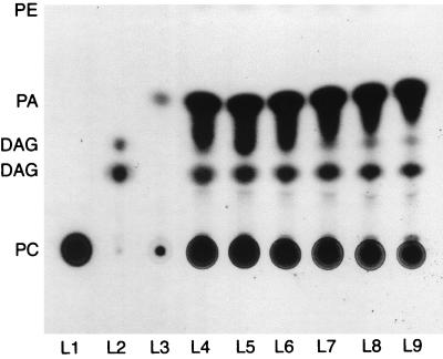
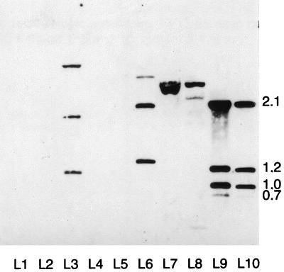
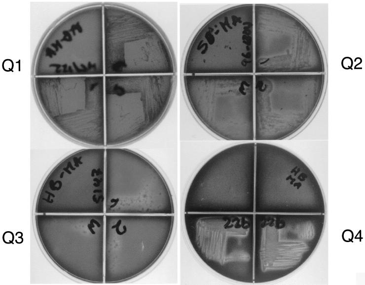
Phospholipase C plays a key role in the pathogenesis of several bacterial infections, for example, those caused by Clostridium perfringens and Listeria monocytogenes. Previous studies have reported multiple copies of plc genes homologous to Pseudomonas aeruginosa plcH and plcN genes encoding the hemolytic and nonhemolytic phospholipase C enzymes in the genomes of Mycobacterium tuberculosis, M. marinum, M. bovis, and M. ulcerans. In this study we analyzed the possible relationship between phospholipase C and hemolytic activity in 21 strains of nontuberculous mycobacteria representing nine different species. Detection of phospholipase C enzymatic activity was carried out using thin-layer chromatography to detect diglycerides in the hydrolysates of radiolabeled phosphatidylcholine. DNA sequences of M. kansasii and M. marinum homologous to the genes encoding phospholipase C from M. tuberculosis and M. ulcerans were identified by DNA-DNA hybridization and sequencing. Finally, we developed a direct and simple assay to detect mycobacterial hemolytic activity. This assay is based on a modified blood agar medium that allows the growth and expression of hemolysis of slow-growing mycobacteria. Hemolytic activity was detected in M. avium, M. intracellulare, M. ulcerans, M. marinum, M. tuberculosis, and M. kansasii mycobacteria with phospholipase C activity, but not in M. fortuitum. No hemolytic activity was detected in M. smegmatis, M. gordonae, and M. vaccae. Whether or not phospholipase C enzyme plays a role in the pathogenesis of nontuberculous mycobacterial diseases needs further investigation.
Figures
Similar articles
-
Biochemical and molecular analysis of phospholipase C and phospholipase D activity in mycobacteria.Infect Immun. 1996 Aug;64(8):3259-66. doi: 10.1128/iai.64.8.3259-3266.1996. Infect Immun. 1996. PMID: 8757862 Free PMC article.
-
Biochemical and genetic evidence for phospholipase C activity in Mycobacterium ulcerans.Infect Immun. 2000 May;68(5):2995-7. doi: 10.1128/IAI.68.5.2995-2997.2000. Infect Immun. 2000. PMID: 10769001 Free PMC article.
-
Nontuberculous mycobacteria: susceptibility pattern and prevalence rate in Shanghai from 2005 to 2008.Chin Med J (Engl). 2010 Jan 20;123(2):184-7. Chin Med J (Engl). 2010. PMID: 20137367
-
Cutaneous Mycobacterial Infections.Clin Microbiol Rev. 2018 Nov 14;32(1):e00069-18. doi: 10.1128/CMR.00069-18. Print 2018 Jan. Clin Microbiol Rev. 2018. PMID: 30429139 Free PMC article. Review.
-
[Bacteriology of mycobacteria: taxonomic and morphological characteristics].Nihon Rinsho. 1998 Dec;56(12):3001-7. Nihon Rinsho. 1998. PMID: 9883600 Review. Japanese.
Cited by
-
Massive gene acquisitions in Mycobacterium indicus pranii provide a perspective on mycobacterial evolution.Nucleic Acids Res. 2012 Nov;40(21):10832-50. doi: 10.1093/nar/gks793. Epub 2012 Sep 10. Nucleic Acids Res. 2012. PMID: 22965120 Free PMC article.
-
Genome structure in the vole bacillus, Mycobacterium microti, a member of the Mycobacterium tuberculosis complex with a low virulence for humans.Microbiology (Reading). 2004 May;150(Pt 5):1519-1527. doi: 10.1099/mic.0.26660-0. Microbiology (Reading). 2004. PMID: 15133113 Free PMC article.
-
Mycobacterium ulcerans cytotoxicity in an adipose cell model.Infect Immun. 2001 Nov;69(11):7182-6. doi: 10.1128/IAI.69.11.7182-7186.2001. Infect Immun. 2001. PMID: 11598099 Free PMC article.
-
Comprehensive profiling of functional attributes, virulence potential and evolutionary dynamics in mycobacterial secretomes.World J Microbiol Biotechnol. 2017 Dec 4;34(1):5. doi: 10.1007/s11274-017-2388-1. World J Microbiol Biotechnol. 2017. PMID: 29204714
-
Roles of Lipolytic enzymes in Mycobacterium tuberculosis pathogenesis.Front Microbiol. 2024 Jan 29;15:1329715. doi: 10.3389/fmicb.2024.1329715. eCollection 2024. Front Microbiol. 2024. PMID: 38357346 Free PMC article. Review.
References
-
- Ahn C H, Lowell J R, Onstad G D, Shuford E H, Hurst G A. A demographic study of disease due to Mycobacterium kansasii or M. intracellulare-avium in Texas. Chest. 1979;75:120–125. - PubMed
-
- Bogner J R, Gathof B, Heinrich B, Matuschke A, Backer U, Goebel F D. Erythrocyte antibodies in AIDS are associated with mycobacteriosis and hypergammaglobulinemia. Klin Wochenschr. 1990;68:1050–1053. - PubMed
-
- Cole S T, Brosch R, Parkhill J, Garnier T, Churcher C, Harris D, Gordon S V, Eiglmeier K, Gas S, Barry C E, III, Tekaia F, Badcock K, Basham D, Brown D, Chillingworth T, Connor R, Davies R, Devlin K, Feltwell T, Gentles S, Hamlin N, Holroyd S, Hornsby T, Jagels K, Barrell B G, et al. Deciphering the biology of Mycobacterium tuberculosis from the complete genome sequence. Nature. 1998;393:537–544. - PubMed
-
- Corbett E L, Churchyard G J, Hay M, Herselman P, Clayton T, Williams B, Hayes R, Mulder D, De Cock K M. The impact of HIV infection on Mycobacterium kansasii disease in South African gold miners. Am J Respir Crit Care Med. 1999;160:10–14. - PubMed
Publication types
MeSH terms
Substances
LinkOut - more resources
Full Text Sources
