

Expression and self-assembly in baculovirus of porcine enteric calicivirus capsids into virus-like particles and their use in an enzyme-linked immunosorbent assay for antibody detection in swine
- PMID: 11283075
- PMCID: PMC87958
- DOI: 10.1128/JCM.39.4.1487-1493.2001
Expression and self-assembly in baculovirus of porcine enteric calicivirus capsids into virus-like particles and their use in an enzyme-linked immunosorbent assay for antibody detection in swine
Abstract
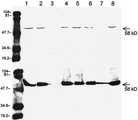
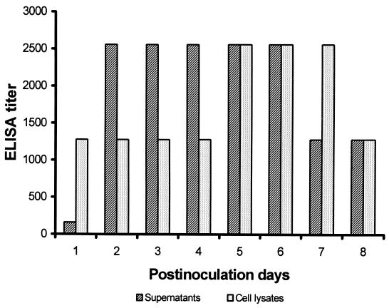
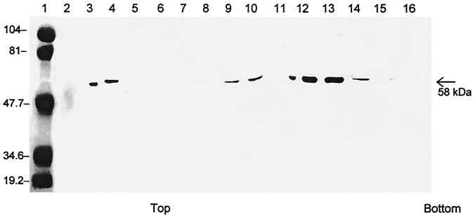
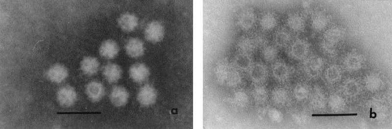
Porcine enteric calicivirus (PEC) causes diarrhea and intestinal lesions in pigs. PEC strain Cowden grows to low to moderate titers in cell culture but only with the addition of intestinal contents from uninfected gnotobiotic pigs (W. T. Flynn and L. J. Saif, J. Clin. Microbiol. 26:206--212, 1988; A. V. Parwani, W. T. Flynn, K. L. Gadfield, and L. J. Saif, Arch. Virol. 120:115--122, 1991). Cloning and sequence analysis of the PEC Cowden full-length genome revealed that it is most closely related genetically to the human Sapporo-like viruses. In this study, the complete PEC capsid gene was subcloned into the plasmid pBlueBac4.5 and the recombinant baculoviruses were identified by plaque assay and PCR. The PEC capsid protein was expressed in insect (Sf9) cells inoculated with the recombinant baculoviruses, and the recombinant capsid proteins self- assembled into virus-like particles (VLPs) that were released into the cell supernatant and purified by CsCl gradient centrifugation. The PEC VLPs had the same molecular mass (58 kDa) as the native virus capsid and reacted with pig hyperimmune and convalescent-phase sera to PEC Cowden in enzyme-linked immunosorbent assay (ELISA) and Western blotting. The PEC capsid VLPs were morphologically and antigenically similar to the native virus by immune electron microscopy. High titers (1:102,400 to 204,800) of PEC-specific antibodies were induced in guinea pigs inoculated with PEC VLPs, suggesting that the VLPs could be useful for future candidate PEC vaccines. A fixed-cell ELISA and VLP ELISA were developed to detect PEC serum antibodies in pigs. For the fixed-cell ELISA, Sf9 cells were infected with recombinant baculoviruses expressing PEC capsids, followed by cell fixation with formalin. For the VLP ELISA, the VLPs were used for the coating antigen. Our data indicate that both tests were rapid, specific, and reproducible and might be used for large-scale serological investigations of PEC antibodies in swine.
Figures
Similar articles
-
Comparative pathogenesis of tissue culture-adapted and wild-type Cowden porcine enteric calicivirus (PEC) in gnotobiotic pigs and induction of diarrhea by intravenous inoculation of wild-type PEC.J Virol. 2001 Oct;75(19):9239-51. doi: 10.1128/JVI.75.19.9239-9251.2001. J Virol. 2001. PMID: 11533186 Free PMC article.
-
Cross-reactivity among sapovirus recombinant capsid proteins.Arch Virol. 2005 Jan;150(1):21-36. doi: 10.1007/s00705-004-0406-8. Epub 2004 Sep 24. Arch Virol. 2005. PMID: 15449145
-
Self-assembly of the recombinant capsid protein of a bovine norovirus (BoNV) into virus-like particles and evaluation of cross-reactivity of BoNV with human noroviruses.J Clin Microbiol. 2005 Feb;43(2):778-85. doi: 10.1128/JCM.43.2.778-785.2005. J Clin Microbiol. 2005. PMID: 15695679 Free PMC article.
-
Porcine enteric caliciviruses: genetic and antigenic relatedness to human caliciviruses, diagnosis and epidemiology.Vaccine. 2007 Jul 26;25(30):5453-66. doi: 10.1016/j.vaccine.2006.12.032. Epub 2006 Dec 29. Vaccine. 2007. PMID: 17234307 Free PMC article. Review.
-
Genetic and antigenic diversity of human caliciviruses (HuCVs) using RT-PCR and new EIAs.Arch Virol Suppl. 1996;12:251-62. doi: 10.1007/978-3-7091-6553-9_27. Arch Virol Suppl. 1996. PMID: 9015122 Review.
Cited by
-
Mutational study of sapovirus expression in insect cells.Virol J. 2005 Feb 23;2:13. doi: 10.1186/1743-422X-2-13. Virol J. 2005. PMID: 15727685 Free PMC article.
-
Reverse genetics system for porcine enteric calicivirus, a prototype sapovirus in the Caliciviridae.J Virol. 2005 Feb;79(3):1409-16. doi: 10.1128/JVI.79.3.1409-1416.2005. J Virol. 2005. PMID: 15650167 Free PMC article.
-
Isolation and characterization of porcine deltacoronavirus from pigs with diarrhea in the United States.J Clin Microbiol. 2015 May;53(5):1537-48. doi: 10.1128/JCM.00031-15. Epub 2015 Mar 4. J Clin Microbiol. 2015. PMID: 25740769 Free PMC article.
-
Comprehensive review of human sapoviruses.Clin Microbiol Rev. 2015 Jan;28(1):32-53. doi: 10.1128/CMR.00011-14. Clin Microbiol Rev. 2015. PMID: 25567221 Free PMC article. Review.
-
Comparative pathogenesis of tissue culture-adapted and wild-type Cowden porcine enteric calicivirus (PEC) in gnotobiotic pigs and induction of diarrhea by intravenous inoculation of wild-type PEC.J Virol. 2001 Oct;75(19):9239-51. doi: 10.1128/JVI.75.19.9239-9251.2001. J Virol. 2001. PMID: 11533186 Free PMC article.
References
-
- Ball J M, Graham D Y, Opekun A R, Estes M K. Recombinant Norwalk virus-like particles given orally to volunteers: phase I study. Gastroenterology. 1999;117:40–48. - PubMed
-
- Bridger J C. Small viruses associated with gastroenteritis in animals. In: Saif L J, Theil K W, editors. Viral diarrheas of man and animals. Boca Raton, Fla: CRC Press; 1990. pp. 161–182.
-
- Dastjerdi A M, Green J, Gallimore C I, Brown D W G, Bridger J C. The bovine Newbury agent-2 is genetically more closely related to human SRSVs than to animal caliciviruses. Virology. 1999;254:1–5. - PubMed
-
- Dingle K E, Lambdem P R, Caul E O, Clarke I N. Human enteric Caliciviridae: the complete genome sequence and expression of virus-like particles from a genetic group II small round structured virus. J Gen Virol. 1995;76:2349–2358. - PubMed
Publication types
MeSH terms
Substances
Grants and funding
LinkOut - more resources
Full Text Sources
Other Literature Sources
