

Case Reports
doi: 10.1128/JCM.39.4.1580-1585.2001.
Isolation of Helicobacter cinaedi from the colon, liver, and mesenteric lymph node of a rhesus monkey with chronic colitis and hepatitis
Affiliations
- PMID: 11283091
- PMCID: PMC87974
- DOI: 10.1128/JCM.39.4.1580-1585.2001
Item in Clipboard
Case Reports
Isolation of Helicobacter cinaedi from the colon, liver, and mesenteric lymph node of a rhesus monkey with chronic colitis and hepatitis
J Clin Microbiol.
2001 Apr.
Abstract
On the basis of biochemical, phenotypic, and 16S rRNA analyses, Helicobacter cinaedi was isolated from the colon, liver, and mesenteric lymph nodes of a 2-year-old rhesus monkey with chronic diarrhea. Histologically, the liver had mild to moderate biliary hyperplasia and hypertrophy with periportal inflammation and fibrosis. Colonic and cecal lesions consisted of diffuse chronic inflammation and glandular hyperplasia extending the length of the crypts. This is the first observation of H. cinaedi associated with active hepatitis and colitis in a nonhuman primate.
Figures
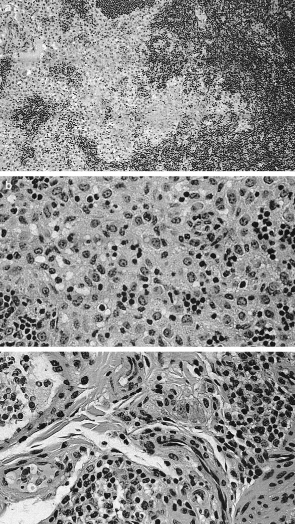
Liver. (a) Portal inflammation with mild peribiliary collagen deposition. (b) Elongated bile duct profiles. (c) Hematoxylin and eosin stain showing centrolobular inflammation. Magnification, ×156 for all panels.
Liver. (a) Portal inflammation with mild peribiliary collagen deposition. (b) Elongated bile duct profiles. (c) Hematoxylin and eosin stain showing centrolobular inflammation. Magnification, ×156 for all panels.
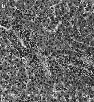
Liver. (a) Portal inflammation with mild peribiliary collagen deposition. (b) Elongated bile duct profiles. (c) Hematoxylin and eosin stain showing centrolobular inflammation. Magnification, ×156 for all panels.
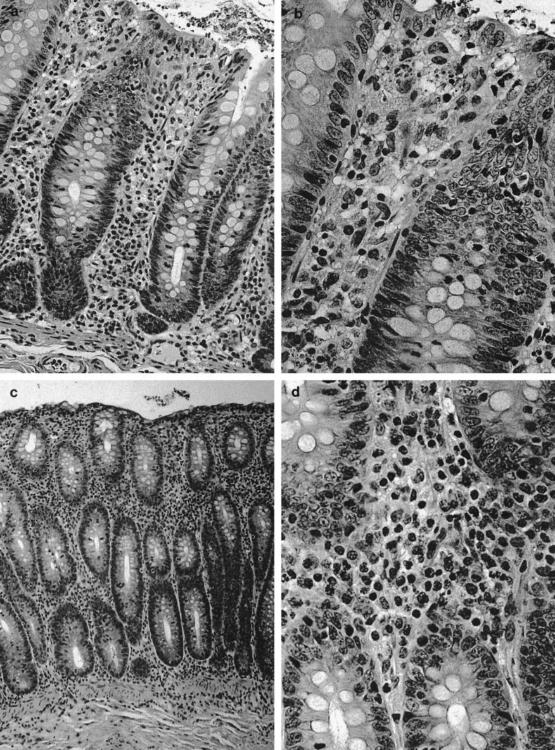
(a and b) Cecal inflammation. Magnifications, ×162 and ×324, respectively. (c and d) Hematoxylin and eosin stain showing colonic inflammation. Magnifications, ×81 and ×324, respectively.
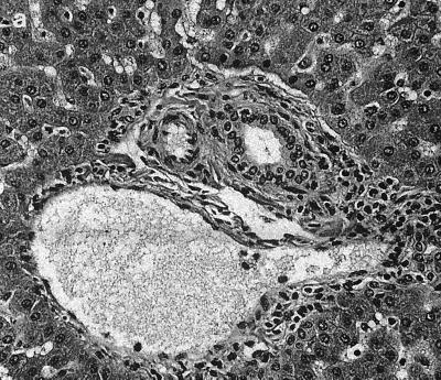
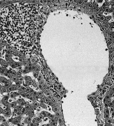
Mesenteric lymph node. (a) Marked hypercellularity of cortex and medulla. Magnification, ×80. (b) Medullary histiocytic infiltrate. Magnification, ×160. (c) Hematoxylin and eosin stain showing inflamed cecal mesentery. Magnification, ×320.
References
-
- Burman W J, Cohn D L, Reves R R, Wilson M L. Multifocal cellulitis and monoarticular arthritis as manifestations of H. cinaedi bacteremia. Clin Infect Dis. 1995;20:564–570. - PubMed
-
- Chalifoux L V, Brieland J K, King N W. Evolution and natural history of colonic disease in cotton-top tamarins (Saguinus oedipus) Dig Dis Sci. 1985;30:54S–58S. - PubMed
Publication types
MeSH terms
Substances
Grants and funding
LinkOut - more resources
Full Text Sources
Medical
Molecular Biology Databases
