. 2001 Apr 2;193(7):855-62.
doi: 10.1084/jem.193.7.855.

CD47-signal regulatory protein alpha (SIRPalpha) regulates Fcgamma and complement receptor-mediated phagocytosis

Affiliations

CD47-signal regulatory protein alpha (SIRPalpha) regulates Fcgamma and complement receptor-mediated phagocytosis

P A Oldenborg et al. J Exp Med. .

Abstract

In autoimmune hemolytic anemia (AIHA), circulating red blood cells (RBCs) opsonized with autoantibody are recognized by macrophage Fcgamma and complement receptors. This triggers phagocytosis and elimination of RBCs from the circulation by splenic macrophages. We recently found that CD47 on unopsonized RBCs binds macrophage signal regulatory protein alpha (SIRPalpha), generating a negative signal that prevents phagocytosis of the unopsonized RBCs. We show here that clearance and phagocytosis of opsonized RBCs is also regulated by CD47-SIRPalpha. The inhibition generated by CD47-SIRPalpha interaction is strongly attenuated but not absent in mice with only residual activity of the phosphatase Src homology 2 domain-containing protein tyrosine phosphatase (SHP)-1, suggesting that most SIRPalpha signaling in this system is mediated by SHP-1 phosphatase activity. The macrophage phagocytic response is controlled by an integration of the inhibitory SIRPalpha signal with prophagocytic signals such as from Fcgamma and complement receptor activation. Thus, augmentation of inhibitory CD47-SIRPalpha signaling may prevent or attenuate RBC clearance in AIHA.


Figures

Figure 1
Figure 1
Clearance of IgG-opsonized RBCs in vivo is regulated by CD47 on the RBCs. Wild-type recipient mice were intravenously injected with PKH26-labeled wild-type RBCs (A; filled symbols) or CD47−/− RBCs (B; open symbols). RBCs were opsonized to different levels with rabbit polyclonal anti–mouse RBCs IgG to obtain low or highly opsonized (ops.) RBCs. Flow cytometric analysis of opsonized RBCs showed identical opsonization of wild-type and CD47−/− RBCs, and a fivefold difference in the level of opsonization between low- (circles) and high-opsonized (squares) RBCs. After extensive washing, 200 μl of low- or high-opsonized RBCs or unopsonized (Unops.) RBCs (triangles; 30% vol/vol in pyrogen-free 0.9% NaCl) was intravenously injected into wild-type recipient mice. Clearance of labeled RBCs was followed using flow cytometry of 5-μl blood samples collected from a tail vein at the time points indicated. Data are mean ± SD for three mice in each group. The rate of clearance increases with increased degree of opsonization and is at each level higher for CD47−/− targets than for CD47+/+ RBCs.
Figure 1
Figure 1
Clearance of IgG-opsonized RBCs in vivo is regulated by CD47 on the RBCs. Wild-type recipient mice were intravenously injected with PKH26-labeled wild-type RBCs (A; filled symbols) or CD47−/− RBCs (B; open symbols). RBCs were opsonized to different levels with rabbit polyclonal anti–mouse RBCs IgG to obtain low or highly opsonized (ops.) RBCs. Flow cytometric analysis of opsonized RBCs showed identical opsonization of wild-type and CD47−/− RBCs, and a fivefold difference in the level of opsonization between low- (circles) and high-opsonized (squares) RBCs. After extensive washing, 200 μl of low- or high-opsonized RBCs or unopsonized (Unops.) RBCs (triangles; 30% vol/vol in pyrogen-free 0.9% NaCl) was intravenously injected into wild-type recipient mice. Clearance of labeled RBCs was followed using flow cytometry of 5-μl blood samples collected from a tail vein at the time points indicated. Data are mean ± SD for three mice in each group. The rate of clearance increases with increased degree of opsonization and is at each level higher for CD47−/− targets than for CD47+/+ RBCs.
Figure 2
Figure 2
Target cell CD47 inhibits Fcγ receptor–mediated phagocytosis of opsonized RBCs in BMMs via ligation of macrophage SIRPα. (A) Effects of anti-SIRPα mAb P84 on phagocytosis of IgG-opsonized RBCs in CD47 wild-type (Wt) BMMs was assayed in medium alone (white bars), in the presence of 10 μg anti-SIRPα mAb P84 (black bars), or in the presence of control mAb anti-CD14 (hatched bars). Wild-type RBCs are phagocytosed at a lower rate than CD47−/− RBCs, which can be virtually completely corrected by anti-SIRPα mAb P84. The rate of CD47−/− RBC phagocytosis can be significantly enhanced by increased opsonization (not shown). Thus, the lack of anti-SIRPα effect on CD47−/− RBC phagocytosis is not due to saturation of the system. (B) Effects of anti-CD47 mAb mIAP301 on phagocytosis of IgG-opsonized wild-type RBCs in wild-type BMMs. Macrophages (Mφ) and RBCs were incubated in medium alone (white bar) or in the presence of 10 μg Fab fragments of anti-CD47 mAb mIAP301 (black bar). To further separate the anti-CD47 blocking effect on macrophage CD47 from that on RBC CD47, respectively, macrophages only (cross-hatched bar) or RBCs only (hatched bar) were preincubated with 10-μg Fab fragments of anti-CD47 mAb mIAP301, extensively washed, and then mixed for assay of phagocytosis. CD47−/− or wild-type RBCs were opsonized to identical levels with rabbit anti–mouse RBC IgG and then added to macrophages (2 × 10⁵) in suspension (100 μl). After 30 min at 37°C, uningested RBCs were lysed and phagocytosis was determined by light microscopy. Results are expressed as number of RBCs ingested per 100 macrophages (phagocytosis index) and are mean ± SEM for three separate experiments. Ligation of target cell CD47 enhances the phagocytosis of wild-type RBCs to the level seen with CD47−/− RBCs.
Figure 3
Figure 3
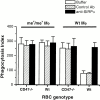
Phagocytosis of C3bi-opsonized RBCs is regulated by target cell CD47 via macrophage SIRPα. BMM (2 × 10⁵) and C3bi-opsonized wild-type (Wt) or CD47−/− RBCs, in the presence of anti-SIRPα mAb P84 (10 μg) or control anti-CD14 mAb (10 μg), were incubated with 30 ng/ml of PDBu and 1 mM MnCl2 in a 100-μl volume for 30 min at 37°C. Uningested RBCs were lysed and phagocytosis was determined by light microscopy. Phagocytosis index is number of RBCs ingested per 100 macrophages and is expressed as mean ± SEM for three separate experiments. To exclude any role of Fcγ receptors, the same experiments were also performed in Fcγ receptor–deficient BMMs.
Figure 4
Figure 4
Fcγ receptor–mediated clearance of IgG-opsonized RBCs is independent of target cell CD47 in SHP-1–deficient motheaten viable (mev/mev) mice. (A) mev/mev or (B) wild-type (Wt) mice were intravenously injected with PKH26-labeled IgG-opsonized wild-type (•) or CD47−/− (▵) RBCs. Opsonization of RBCs and clearance of injected RBCs was performed and followed as described in the legend to Fig. 1. Data are mean ± SD for three mice in each group.
Figure 5
Figure 5
Phagocytosis of IgG-opsonized RBCs is independent of CD47 in BMMs from SHP-1–deficient mev/mev mice. BMMs (Mφ) from mev/mev or wild-type (Wt) mice were incubated with IgG-opsonized RBCs in medium alone (white bars), in the presence of 10 μg anti-SIRPα mAb P84 (black bars) or control anti-CD14 mAb (hatched bars). Phagocytosis was determined as described in the legend to Fig. 2. Data are presented as mean ± SEM for three separate experiments.
Figure 6
Figure 6
Residual and attenuated inhibitory CD47-SIRPα signal in mev/mev mice. Clearance of unopsonized PKH26-labeled CD47+/+ (•), CD47+/− (▪), or CD47−/− (▵) RBCs in (A) mev/mev or (B) wild-type (Wt) mice. Clearance of unopsonized RBCs was performed as described in the legend to Fig. 1. Data are mean ± SD for three to five mice in each group.
Figure 7
Figure 7
A model for the regulation of macrophage phagocytosis based on a summation of positive prophagocytic and negative CD47-SIRPα–generated signals. Neither type of signal is dominant. Rather, the decision to phagocytose is based on the summation of weak positive signals (the RBC receptor), strong positive signals (Fc and complement receptors), and negative signals (SIRPα). The majority of SIRPα signaling is mediated via SHP-1 as shown by the marked reduction of the signal in mev/mev mice. The data presented do not rule out the possibility that a small subset of the SIRPα signal may be mediated by SHP-2.

References

    1. Clynes R., Ravaetch J.V. Cytotoxic antibodies trigger inflammation through Fc receptors. Immunity. 1995;3:21–26. - PubMed
    1. Meyer D., Schiller C., Westermann J., Izui S., Hazenbos W.L.W., Verbeek J.S., Schmidt R.E., Gessner J.E. FcγRIII (CD16)-deficient mice show IgG isotype-dependent protection to experimental autoimmune hemolytic anemia. Blood. 1998;92:3997–4002. - PubMed
    1. Schreiber A.D., Frank M.M. Role of antibody and complement in the immune clearance and destruction of erythrocytes. I. In vivo effects of IgG and IgM complement fixing sites. J. Clin. Invest. 1972;51:575–582. - PMC - PubMed
    1. Schreiber A.D., Frank M.M. Role of antibody and complement in the immune clearance and destruction of erythrocytes. II. Molecular nature of IgG and IgM complement-fixing sites and effects of their interaction with serum. J. Clin. Invest. 1972;51:583–589. - PMC - PubMed
    1. Oldenborg P.-A., Zheleznyak A., Fang Y.-F., Lagenaur C.F., Gresham H.D., Lindberg F.P. Role of CD47 as a marker of self on red blood cells. Science. 2000;288:2051–2054. - PubMed

Publication types

MeSH terms