. 2001 Apr;21(8):2695-705.
doi: 10.1128/MCB.21.8.2695-2705.2001.

Sustained signaling by phospholipase C-gamma mediates nerve growth factor-triggered gene expression

Affiliations

Sustained signaling by phospholipase C-gamma mediates nerve growth factor-triggered gene expression

D Y Choi et al. Mol Cell Biol. 2001 Apr.

Abstract

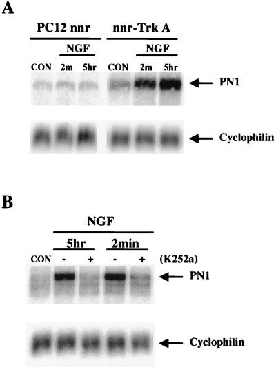
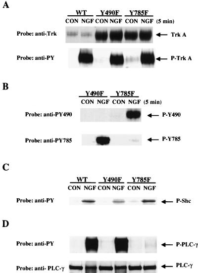
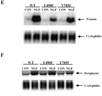
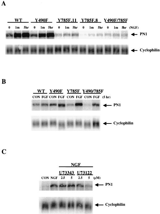
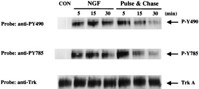
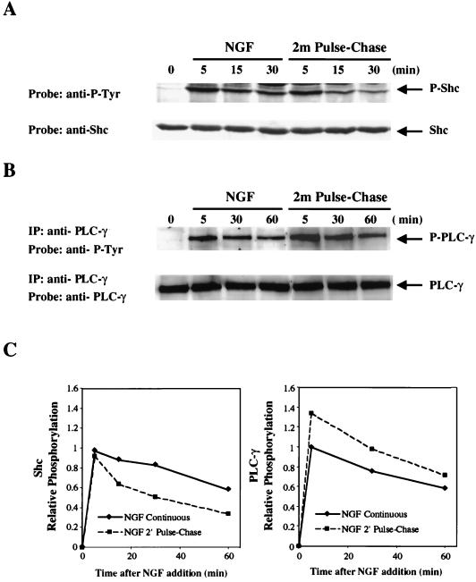
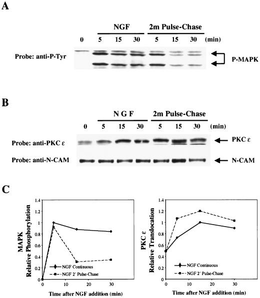
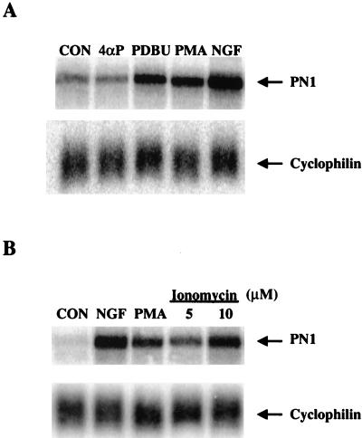
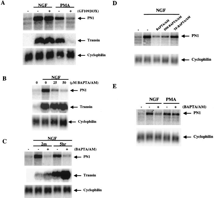
In contrast to conventional signaling by growth factors that requires their continual presence, a 1-min pulse of nerve growth factor (NGF) is sufficient to induce electrical excitability in PC12 cells due to induction of the peripheral nerve type 1 (PN1) sodium channel gene. We have investigated the mechanism for this triggered signaling pathway by NGF in PC12 cells. Mutation of TrkA at key autophosphorylation sites indicates an essential role for the phospholipase C-gamma (PLC-gamma) binding site, but not the Shc binding site, for NGF-triggered induction of PN1. In concordance with results with Trk mutants, drug-mediated inhibition of PLC-gamma activity also blocks PN1 induction by NGF. Examination of the kinetics of TrkA autophosphorylation indicates that triggered signaling does not result from sustained activation and autophosphorylation of the TrkA receptor kinase, whose phosphorylation state declines rapidly after NGF removal. Rather, TrkA triggers an unexpectedly prolonged phosphorylation and activation of PLC-gamma signaling that is sustained for up to 2 h. Prevention of the elevation of intracellular Ca2+ levels using BAPTA-AM results in a block of PN1 induction by NGF. Sustained signaling by PLC-gamma provides a means for differential neuronal gene induction after transient exposure to NGF.


Figures

FIG. 1
FIG. 1
TrkA tyrosine kinase activity is required for NGF induction of PN1. (A) Total cellular RNA (10 μg) was isolated from PC12 nnr5 (lacking TrkA) and nnr5-TrkA (T14 cells) cells expressing wild-type TrkA, at 5 h after either a 2-min pulsatile treatment or continuous treatment with 50 ng of NGF per ml. RNA was electrophoresed through a 0.8% agarose gel and blotted. Blots were hybridized with a sodium channel probe (top) and rehybridized with a probe specific for the internal control cyclophilin (bottom). (B) PC12 cells were pretreated with 150 nM K252a for 20 min and then treated with 50 ng of NGF per ml for 5 h. Northern blots were prepared and analyzed as described in the legend to panel A.
FIG. 2
FIG. 2
Specific signaling defects of TrkA mutants. (A) PC12 nnr5 cells expressing wild-type TrkA, TrkA Y490F, and TrkA Y785F were treated with 50 ng of NGF per ml for 5 min. Cell lysates were electrophoresed through a SDS–7.5% polyacrylamide gel and blotted onto a nitrocellulose filter. The blot was probed with anti-TrkA antibody (top) and an anti-phosphotyrosine (4G10) antibody (bottom). (B) PC12 nnr5 Y490F and Y785F cells were treated with 50 ng of NGF per ml for 5 min. Cell lysates were electrophoresed and blotted onto nitrocellulose. The blots were probed with antibodies specific for individual phosphotyrosine residues of TrkA (anti-P-Y490 or anti-P-Y785). (C) PC12 nnr5 wild-type (WT), Y490F, and Y785F cells were treated with 50 ng of NGF per ml for 5 min. Whole-cell lysates were electrophoresed and blotted, and the blots were probed with anti-phosphotyrosine antibody. (D) PC12 nnr5 WT, Y490F, and Y785F cells were treated with 50 ng of NGF per ml for 5 min. PLC-γ was immunoprecipitated from cell lysates using anti-PLC-γ antibody. Immunoprecipitates were electrophoresed and blotted onto nitrocellulose. The blots were probed with anti-phosphotyrosine antibody (top). The blots were then stripped and reprobed with anti-PLC-γ antibody (bottom). (E) PC12 nnr5 WT, Y490F, and Y785F cells were treated with 50 ng of NGF per ml for 5 h. Total cellular RNA (10 μg) was isolated and electrophoresed through 0.8% agarose gels and blotted. Blots were hybridized with a probe specific for transin (1.9 kb) (top). The blots were rehybridized with a probe specific for cyclophilin (1 kb) as an internal standard (bottom). (F) Cells were treated with 50 ng of NGF per ml for 48 h. Total cellular RNA (10 μg) was isolated and electrophoresed through 1.4% agarose gels and blotted. The blot was hybridized with a probe specific for peripherin (top) or with a probe specific for the internal control cyclophilin (bottom).
FIG. 2
FIG. 2
Specific signaling defects of TrkA mutants. (A) PC12 nnr5 cells expressing wild-type TrkA, TrkA Y490F, and TrkA Y785F were treated with 50 ng of NGF per ml for 5 min. Cell lysates were electrophoresed through a SDS–7.5% polyacrylamide gel and blotted onto a nitrocellulose filter. The blot was probed with anti-TrkA antibody (top) and an anti-phosphotyrosine (4G10) antibody (bottom). (B) PC12 nnr5 Y490F and Y785F cells were treated with 50 ng of NGF per ml for 5 min. Cell lysates were electrophoresed and blotted onto nitrocellulose. The blots were probed with antibodies specific for individual phosphotyrosine residues of TrkA (anti-P-Y490 or anti-P-Y785). (C) PC12 nnr5 wild-type (WT), Y490F, and Y785F cells were treated with 50 ng of NGF per ml for 5 min. Whole-cell lysates were electrophoresed and blotted, and the blots were probed with anti-phosphotyrosine antibody. (D) PC12 nnr5 WT, Y490F, and Y785F cells were treated with 50 ng of NGF per ml for 5 min. PLC-γ was immunoprecipitated from cell lysates using anti-PLC-γ antibody. Immunoprecipitates were electrophoresed and blotted onto nitrocellulose. The blots were probed with anti-phosphotyrosine antibody (top). The blots were then stripped and reprobed with anti-PLC-γ antibody (bottom). (E) PC12 nnr5 WT, Y490F, and Y785F cells were treated with 50 ng of NGF per ml for 5 h. Total cellular RNA (10 μg) was isolated and electrophoresed through 0.8% agarose gels and blotted. Blots were hybridized with a probe specific for transin (1.9 kb) (top). The blots were rehybridized with a probe specific for cyclophilin (1 kb) as an internal standard (bottom). (F) Cells were treated with 50 ng of NGF per ml for 48 h. Total cellular RNA (10 μg) was isolated and electrophoresed through 1.4% agarose gels and blotted. The blot was hybridized with a probe specific for peripherin (top) or with a probe specific for the internal control cyclophilin (bottom).
FIG. 3
FIG. 3
NGF-induced PN1 expression is specifically blocked in cells expressing TrkA Y785F. (A) Total cellular RNA (10 μg) was isolated from PC12 nnr5 wild-type (WT), Y490F, Y785F, and Trk Y490F/785F cells at 5 h after either a 1-min pulsatile or continuous treatment with 50 ng of NGF per ml. RNA was electrophoresed and blotted, and the blots were probed with a sodium channel probe, pRB211, and rehybridized with a cyclophilin probe, p1B15. (B) Total cellular RNA (10 μg) was isolated from PC12 nnr5 WT, Y490F, Y785F, and Trk Y490F-785F cells at 5 h after treatment with 50 ng of FGF-1 per ml. RNA was hybridized with a sodium channel probe, pRB211 (top), and rehybridized with a probe specific for cyclophilin (bottom). (C) Northern blots prepared from total cellular RNA (10 μg) isolated from PC12 cells at 5 h after treatment with indicated amounts of U73343, U73122, and/or 50 ng of NGF per ml (cells were also pretreated with the inhibitors for 30 min before NGF treatment) were hybridized with a sodium channel probe (top) and rehybridized with a cyclophilin probe for an internal control (bottom).
FIG. 4
FIG. 4
Both Y785 and Y490 sites are transiently phosphorylated after pulsatile NGF treatment. Protein extracts were prepared from TrkA PC12 cells at the indicated times after either a 2-min pulsatile (pulse-chase) treatment or continuous treatment with 50 ng of NGF per ml. The proteins were electrophoresed through a SDS–7.5% polyacrylamide gel and blotted onto nitrocellulose. The blot was probed with anti-P-Y490 antibody (top). A parallel blot made from the same samples was probed with anti-P-Y785 antibody (middle). After stripping, the blots were reprobed with anti-TrkA antibody (bottom).
FIG. 5
FIG. 5
Pulsatile NGF treatment triggers prolonged tyrosine phosphorylation of PLC-γ, but not of Shc. (A) Cell lysates were prepared from PC12 cells at the indicated times after either 2 min of pulsatile (pulse-chase) treatment or continuous treatment with 50 ng of NGF per ml. The proteins were electrophoresed and blotted onto nitrocellulose, and the blots were probed with anti-phosphotyrosine antibody (anti-P-Tyr) (top). After being stripped, the blot was probed again with anti-Shc antibody (bottom). (B) PLC-γ was immunoprecipitated from the cell lysates with anti-PLC-γ antibody. Western blots of the imunoprecipitated proteins were probed with anti-phosphotyrosine antibody (top). After being stripped, the blot was probed with anti-PLC-γ antibody (bottom). (C) Quantification was performed using a densitometer and software as described in Materials and Methods. The maximal response to continuous NGF treatment was given a value of 1.0.
FIG. 6
FIG. 6
Pulsatile NGF treatment triggers prolonged activation of protein kinase C, but not of MAPK. (A) Cell lysates were prepared from PC12 cells at the indicated times after either 2 min of pulsatile (pulse-chase) treatment or continuous treatment with 50 ng of NGF per ml. The proteins were electrophoresed and blotted onto nitrocellulose, and the blots were probed with anti-phosphotyrosine antibody. The positions of the ERK1 and ERK2 MAPKs are indicated. (B) At the indicated times after either 2 min of pulsatile (pulse-chase) treatment or continuous treatment with 50 ng of NGF per ml, PC12 cells were fractionated into cytosolic and membrane fractions by sonication and subsequent centrifugation. Membrane proteins were extracted, electrophoresed, and blotted, and the blots were probed with anti-protein kinase C-ɛ (anti-PKC ɛ) antibody (top) and with anti-N-CAM antibody (bottom). (C) Quantification was performed as described in Materials and Methods. The maximal response to continuous NGF treatment was given a value of 1.0.
FIG. 7
FIG. 7
PN1 mRNA expression is induced by both protein kinase C activators and calcium ionophore. (A) Northern blots prepared from total cellular RNA (10 μg) isolated from PC12 cells at 5 h after treatment with 0.1 μM 4-α-phorbol, PMA, PDBU or 50 ng of NGF per ml were hybridized with a sodium channel probe (top) and rehybridized with cyclophilin probe for an internal control (bottom). (B) Northern blots prepared from total cellular RNA (10 μg) isolated from PC12 cells at 5 h after treatment with PMA, ionomycin, or 50 ng of NGF per ml were hybridized with a sodium channel probe (top) and rehybridized with cyclophilin probe for an internal control (bottom).
FIG. 8
FIG. 8
Effects of the protein kinase C-specific inhibitor, GF109203X, and the intracellular calcium chelator, BAPTA-AM, on PN1 gene expression mediated by PMA and NGF. (A) PC12 cells were pretreated with 1 μM GF109203X for 1 h and then treated with 50 ng of NGF per ml or 0.1 μM PMA for 5 h. Northern blots of total cellular RNA (10 μg) were probed with a sodium channel probe (top) and rehybridized with a probe specific for transin (middle) and a probe specific for cyclophilin (bottom). (B) PC12 cells were pretreated with 25 or 50 μM BAPTA-AM for 1 h and then treated for 5 h with 50 ng of NGF per ml. Northern blots of total cellular RNA (10 μg) were probed with a sodium channel probe pRB211 (top) and rehybridized with a probe specific for transin (middle) and a probe specific for cyclophilin (bottom). (C) PC12 cells were pretreated with 50 μM BAPTA-AM for 1 h and then treated for 5 h with 50 ng of NGF per ml or for 2 min with NGF and incubated for a total of 5 h. Northern blots of total cellular RNA (10 μg) were probed with a sodium channel probe (top) and rehybridized with a probe specific for transin (middle) and a probe specific for cyclophilin (bottom). (D) PC12 cells were pretreated with 40 μM BAPTA-AM, dimethyl(DM) BAPTA-AM, or tetraflouro(TF)-BAPTA-AM for 1 h and then treated for 5 h with 50 ng of NGF per ml. Northern blots of total cellular RNA (10 μg) were probed with a sodium channel probe (top) and rehybridized with a probe specific for cyclophilin (bottom). (E) PC12 cells were pretreated with 40 μM BAPTA-AM for 1 h and then treated for 5 h with 50 ng of NGF per ml or 0.1 μM PMA. Northern blots of total cellular RNA (10 μg) were probed with a sodium channel probe (top) and rehybridized with a probe specific for cyclophilin (bottom).

Similar articles

Cited by

References

    1. Altar C A, DiStefano P S. Neurotrophin trafficking by anterograde transport. Trends Neurosci. 1998;21:433–437. - PubMed
    1. Barbacid M. Neurotrophic factors and their receptors. Curr Opin Cell Biol. 1995;7:148–155. - PubMed
    1. Berg M M, Sternberg D W, Parada L F, Chao M V. K-252a inhibits nerve growth factor-induced trk proto-oncogene tyrosine phosphorylation and kinase activity. J Biol Chem. 1992;267:13–16. - PubMed
    1. Bonni A, Ginty D D, Dudek H, Greenberg M E. Serine 133-phosphorylated CREB induces transcription via a cooperative mechanism that may confer specificity to neurotrophin signals. Mol Cell Neurosci. 1995;6:168–183. - PubMed
    1. Bourne H R, Sanders D A, McCormick F. The GTPase superfamily: a conserved switch for diverse cell functions. Nature. 1990;348:125–132. - PubMed

Publication types

MeSH terms

LinkOut - more resources