. 2001 Apr;21(8):2716-25.
doi: 10.1128/MCB.21.8.2716-2725.2001.

Structure and specificity of GATA proteins in Th2 development

Affiliations

Structure and specificity of GATA proteins in Th2 development

S Ranganath et al. Mol Cell Biol. 2001 Apr.

Abstract

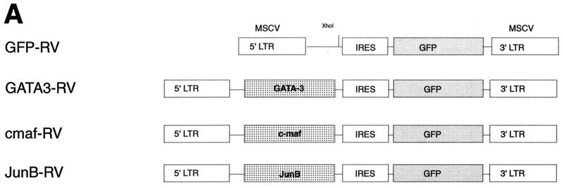
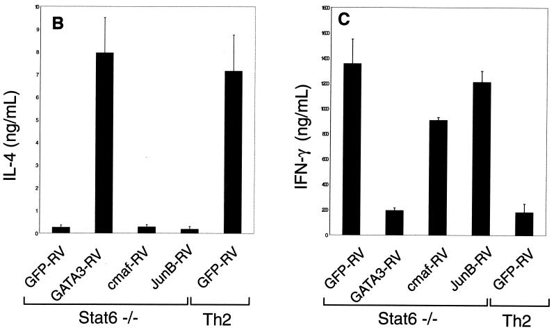
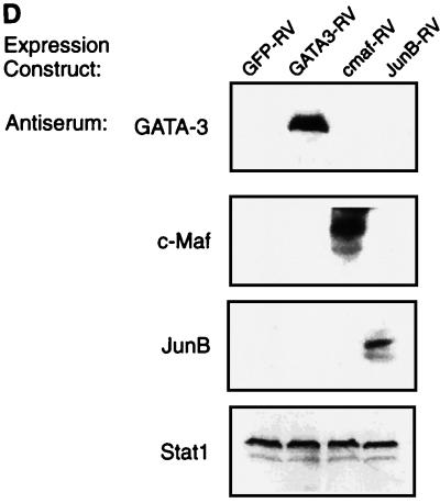
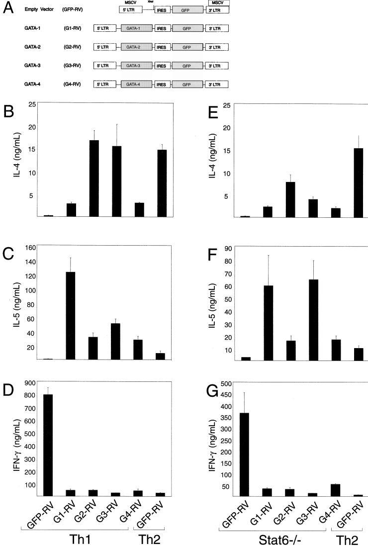
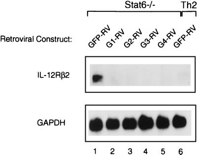
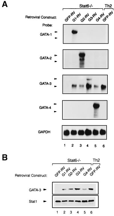
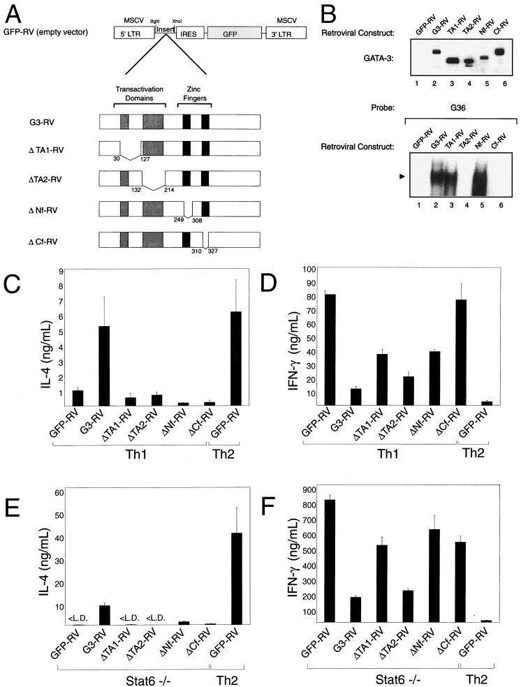
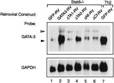
Development of Th2 subset of CD4+ T cells involves the interleukin-4 (IL-4)- and Stat6-dependent increase in GATA-3 expression during primary activation. Recently we reported that the phenotypic stability and factor independence of Th2 cells involves acquisition of an intracellular pathway that maintains GATA-3 expression. Evidence from retroviral expression studies implied that this pathway involved an autoactivation of GATA-3 expression, since Stat6-deficient T cells induced endogenous GATA-3 when infected with GATA-3-expressing retroviruses. That study left unresolved the issue of whether GATA-3 autoactivation was direct or indirect. Several other Th2-specific transcription factors have been described, including c-Maf and JunB. We therefore examined the ability of these other transcription factors to induce GATA-3 expression and promote Th2 development. Neither c-Maf nor JunB induced Th2 development in Stat6-deficient CD4+ T cells, in contrast to GATA-3. Consistent with this indication of a possible direct autoactivation pathway, we also observed that heterologous GATA family proteins GATA-1, GATA-2, and GATA-4 were also capable of inducing GATA-3 expression in developing Stat6-deficient T cells and promote Th2 development. Mutational analysis revealed evidence for two distinct mechanisms of GATA-3 action. IL-4 induction by GATA-3 required each of the functional domains to be present, whereas repression of gamma interferon could occur even when mutants of GATA-3 lacking the second transactivation domain, TA2, were expressed. The GATA-dependent induction of the GATA-3 but not the other GATA genes in T cells suggests that T-cell-specific cis elements within the GATA-3 locus likely cooperate with a general GATA recognition motif to allow GATA-3-dependent autoactivation.


Figures

FIG. 1
FIG. 1
GATA-3 but not c-Maf or JunB induces IL-4 expression in Stat6-deficient T cells. (A) Schematic of GFP-RV-based retroviral expression constructs containing GATA-3 (GATA3-RV), c-Maf (cmaf-RV), or JunB (JunB-RV). (B) Induction of IL-4 by GATA-3 but not by c-Maf or JunB in Stat6-deficient T cells. Activated Stat6-deficient or wild-type DO11.10 splenocytes developed under Th2 conditions were transduced with the indicated retrovirus as described (39). Cells were sorted 7 days after the first activation, restimulated with ovalbumin (0.5 mg/ml) and irradiated BALB/c splenocytes (2.5 × 10⁶ cells, 2,000 rad) for 1 week under the initial conditions, then harvested (day 14), and restimulated. Supernatants were collected after 48 h (day 16) for IL-4 ELISA analysis. Data bars represent the average of four independent cytokine readings for each point, and error bars show the standard deviation. (C) c-Maf and JunB fail to suppress production of IFN-γ in Stat6-deficient T cells. Supernatants described in for panel B were subjected to quantitation of IFN-γ levels by ELISA as described (14). Data are presented as in panel B. (D) Expression of murine stem cell virus (MSCV) long terminal repeat (LTR)-driven GATA-3, c-Maf, and JunB. 293 cells were transiently transfected with 20 μg of GATA3-RV, cmaf-RV, and JunB-RV using Superfect (Quiagen). After 48 h, cells lysates were electrophoresed by SDS–12% PAGE, transferred to nitrocellulose, and analyzed by Western analysis. GATA-3 was detected using antibody HG3-31 (Santa Cruz), c-Maf was detected using M173 (Santa Cruz), JunB was detected using N17 (Santa Cruz), and Stat1 was detected using E23 (Santa Cruz).
FIG. 1
FIG. 1
GATA-3 but not c-Maf or JunB induces IL-4 expression in Stat6-deficient T cells. (A) Schematic of GFP-RV-based retroviral expression constructs containing GATA-3 (GATA3-RV), c-Maf (cmaf-RV), or JunB (JunB-RV). (B) Induction of IL-4 by GATA-3 but not by c-Maf or JunB in Stat6-deficient T cells. Activated Stat6-deficient or wild-type DO11.10 splenocytes developed under Th2 conditions were transduced with the indicated retrovirus as described (39). Cells were sorted 7 days after the first activation, restimulated with ovalbumin (0.5 mg/ml) and irradiated BALB/c splenocytes (2.5 × 10⁶ cells, 2,000 rad) for 1 week under the initial conditions, then harvested (day 14), and restimulated. Supernatants were collected after 48 h (day 16) for IL-4 ELISA analysis. Data bars represent the average of four independent cytokine readings for each point, and error bars show the standard deviation. (C) c-Maf and JunB fail to suppress production of IFN-γ in Stat6-deficient T cells. Supernatants described in for panel B were subjected to quantitation of IFN-γ levels by ELISA as described (14). Data are presented as in panel B. (D) Expression of murine stem cell virus (MSCV) long terminal repeat (LTR)-driven GATA-3, c-Maf, and JunB. 293 cells were transiently transfected with 20 μg of GATA3-RV, cmaf-RV, and JunB-RV using Superfect (Quiagen). After 48 h, cells lysates were electrophoresed by SDS–12% PAGE, transferred to nitrocellulose, and analyzed by Western analysis. GATA-3 was detected using antibody HG3-31 (Santa Cruz), c-Maf was detected using M173 (Santa Cruz), JunB was detected using N17 (Santa Cruz), and Stat1 was detected using E23 (Santa Cruz).
FIG. 2
FIG. 2
Heterologous GATA factors induce Th2 development. (A) Schematic of constructs driving the expression of control vector (GFP-RV), GATA-1 (G1-RV), GATA2 (G2-RV), GATA-3 (G3-RV), and GATA-4 (G4-RV). IL-4 (B and E) and IL-5 (C and F) induction by GATA-1, GATA-2, GATA-3, and GATA-4 in developing DO11.10 Th1 cells and Stat6-deficient T cells. Activated splenocytes were developed under the indicated conditions and transduced with the indicated retroviral expression constructs 36 h postactivation. Cells were sorted 7 days after the first activation for expression of GFP and murine CD4 to greater than 95% puarity, restimulated with ovalbumin protein (0.5 mg/ml) and irradiated BALB/c splenocytes (2.5 × 10⁶ cells, 2,000 rad) for 1 week under the initial conditions, harvested on day 14, and restimulated for cytokine analysis. Supernatants were collected after 48 h (day 16) and subjected to IL-4 and IL-5 ELISA analysis. Data are presented as in Fig. 1B. (D and G) GATA-1, GATA-2, GATA-3, and GATA-4 repress IFN-γ production by developing wild-type Th1 cells (D) and Stat6-deficient cells (G). The supernatants collected from the cells described above were quantified for their levels of IFN-γ by ELISA. Data are presented as in Fig. 1B.
FIG. 3
FIG. 3
Repression of IL-12Rβ2 expression by heterologous GATA factors. Northern analysis of IL-12Rβ2 mRNA in cells transduced with GATA-1, GATA-2, GATA-3, and GATA-4. Stat6-deficient lymphocytes transduced with GATA proteins (as described in the legend to Fig. 2E) were restimulated and expanded for subsequent Northern analysis. Cells were harvested on day 28 and restimulated with PMA (50 ng/ml) and ionomycin (1 M) for 6 h prior to RNA isolation; 10 μg of total RNA was subjected to electrophoresis on a 1.1% agarose gel, transferred to Zeta-probe membrane, and probed sequentially with IL-12Rβ2 and GAPDH probes as described (43).
FIG. 4
FIG. 4
Heterologous GATA family proteins induce endogenous GATA-3 expression. (A) Northern analysis of retroviral and endogenous mRNA in cells transduced with GATA-1, GATA-2, GATA-3, and GATA-4. The cells described in the legend to Fig. 2E were analyzed for expression of GATA-1, GATA-2, GATA-3, and GATA-4 as described for Fig. 3. Open triangles indicate the predicted retroviral GATA mRNA, and solid triangles indicate the predicted endogenous GATA mRNA. (B) Western analysis of GATA-3 protein levels. Cells were harvested on day 28 and stimulated with PMA (50 ng/ml) and ionomycin (1 μM) for 16 h. Cytoplasmic and nuclear proteins were extracted as described in Materials and Methods, then electrophoresed through an SDS–12% PAGE gel, and transferred to nitrocellulose. GATA-3 was detected using antibody HG3-31, and Stat1 was detected using antibody E23 (Santa Cruz) as a normalization control.
FIG. 5
FIG. 5
Distinct domains couple GATA-3 to IL-4 activation and IFN-γ inhibition. (A) Schematic of GATA-3 mutations, illustrating the four described functional domains of GATA-3 and the amino acids (aa) deleted from each specific expression construct: ΔTA1-RV, deletion of GATA-3 residues 29 to 128; ΔTA2-RV, deletion of GATA-3 132 to 214; ΔNf, deletion of GATA-3 249 to 308, encompassing the entire N-terminal zinc finger; and ΔCf, deletion of GATA-3 309 to 328, a portion of the C-terminal zinc finger region. (B) QT6 cells were transiently transfected with the indicated GATA-3 expression constructs, and nuclear extracts were prepared after 48 h. Nuclear extracts were electrophoresed by SDS–12% PAGE, transferred to nitrocellulose, and probed for GATA-3 expression (upper panel). Nuclear extracts were incubated in 10-μl reactions with the GATA-3 probe for 30 min and resolved by 6% polyacrylamide electrophoresis at room temperature. The solid triangle indicates the probe-bound GATA complex. (C and E) Mutant GATA proteins fail to activate IL-4 in developing Th1 and Stat6-deficient DO11.10 cells. Data are presented as in Fig. 1B. (D and F) Mutant GATA proteins differentially repress IFN-γ production in developing Th1 (D) and Stat6-deficient (F) T cells. Data are presented as in Fig. 1B. L.D., limit of detection.
FIG. 6
FIG. 6
Structural requirements for GATA-3 autoactivation. Cells described in the legends to Fig. 5D and 5E were harvested on day 28, and total RNA was isolated; 10 μg of total RNA was subjected to electrophoresis on a 1.1% agarose gel, transferred to Zeta-probe membrane, and probed sequentially with GATA-3 (35) and GAPDH (43) probes as described. The open triangle indicates the predicted retroviral GATA-3 mRNA, and the solid triangle indicates the predicted endogenous GATA-3 mRNA.

Similar articles

Cited by

References

    1. Arceci R J, King A A, Simon M C, Orkin S H, Wilson D B. Mouse GATA-4: a retinoic acid-inducible GATA-binding transcription factor expressed in endodermally derived tissues and heart. Mol Cell Biol. 1993;13:2235–2246. - PMC - PubMed
    1. Blobel G A, Nakajima T, Eckner R, Montminy M, Orkin S H. CREB-binding protein cooperates with transcription factor GATA-1 and is required for erythroid differentiation. Proc Natl Acad Sci USA. 1998;95:2061–2066. - PMC - PubMed
    1. Boyes J, Byfield P, Nakatani Y, Ogryzko V. Regulation of activity of the transcription factor GATA-1 by acetylation. Nature. 1998;396:594–598. - PubMed
    1. Dasen J S, O'Connell S M, Flynn S E, Treier M, Gleiberman A S, Szeto D P, Hooshmand F, Aggarwal A K, Rosenfeld M G. Reciprocal interactions of Pit1 and GATA2 mediate signaling gradient-induced determination of pituitary cell types. Cell. 1999;97:587–598. - PubMed
    1. Dorfman D M, Wilson D B, Bruns G A, Orkin S H. Human transcription factor GATA-2: evidence for regulation of preproendothelin-1 gene expression in endothelial cells. J Biol Chem. 1992;267:1279–1285. - PubMed

Publication types

MeSH terms