. 2001 Apr;21(8):2755-66.
doi: 10.1128/MCB.21.8.2755-2766.2001.

S and G2 phase roles for Cdk2 revealed by inducible expression of a dominant-negative mutant in human cells

Affiliations

S and G2 phase roles for Cdk2 revealed by inducible expression of a dominant-negative mutant in human cells

B Hu et al. Mol Cell Biol. 2001 Apr.

Abstract

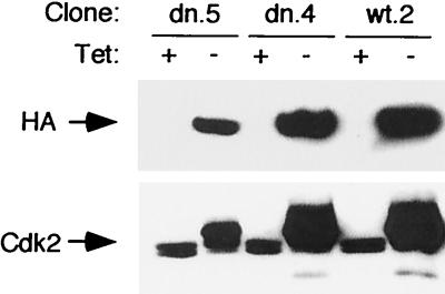
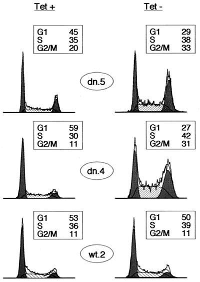
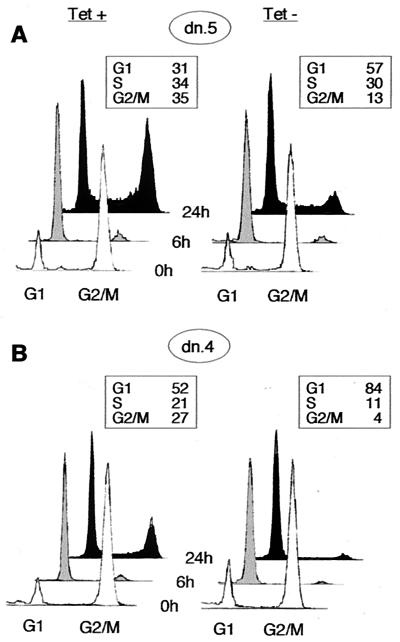
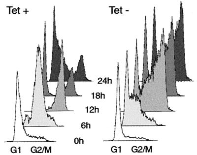
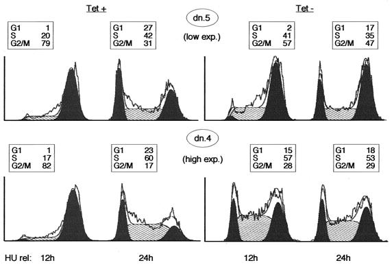
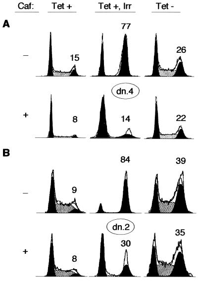
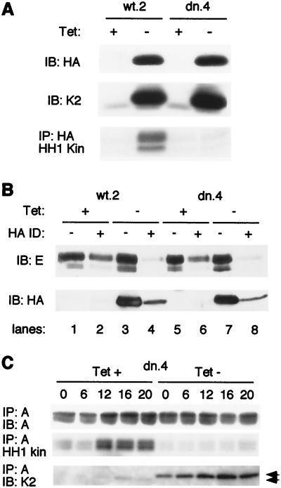
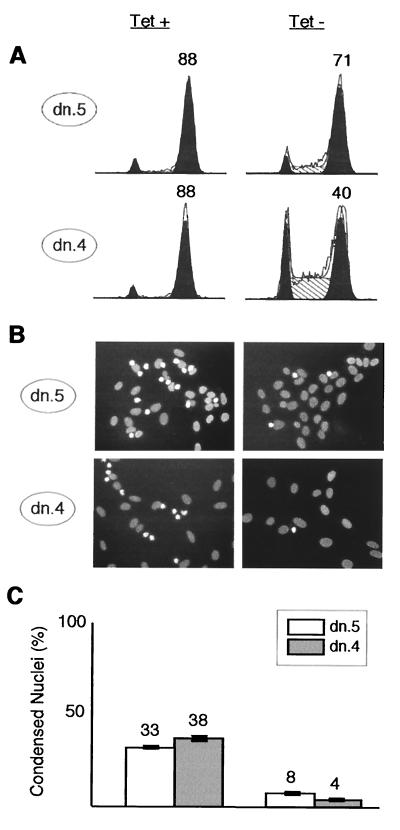
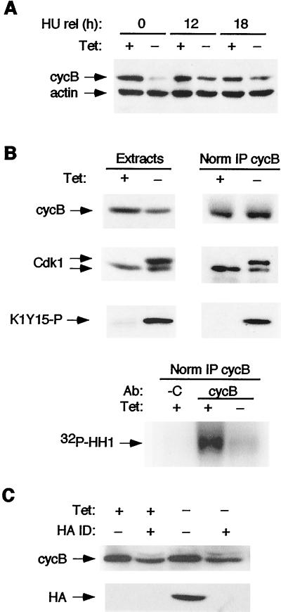
Cyclin-dependent kinase 2 (Cdk2) is essential for initiation of DNA synthesis in higher eukaryotes. Biochemical studies in Xenopus egg extracts and microinjection studies in human cells have suggested an additional function for Cdk2 in activation of Cdk1 and entry into mitosis. To further examine the role of Cdk2 in human cells, we generated stable clones with inducible expression of wild-type and dominant-negative forms of the enzyme (Cdk2-wt and Cdk2-dn, respectively). Both exogenous proteins associated efficiently with endogenous cyclins. Cdk2-wt had no apparent effect on the cell division cycle, whereas Cdk2-dn inhibited progression through several distinct stages. Cdk2-dn induction could arrest cells at the G1/S transition, as previously observed in transient expression studies. However, under normal culture conditions, Cdk2-dn induction primarily arrested cells with S and G2/M DNA contents. Several observations suggested that the latter cells were in G2 phase, prior to the onset of mitosis: these cells contained uncondensed chromosomes, low levels of cyclin B-associated kinase activity, and high levels of tyrosine-phosphorylated Cdk1. Furthermore, Cdk2-dn did not delay progression through mitosis upon release of cells from a nocodazole block. Although the G2 arrest imposed by Cdk2-dn was similar to that imposed by the DNA damage checkpoint, the former was distinguished by its resistance to caffeine. These findings provide evidence for essential functions of Cdk2 during S and G2 phases of the mammalian cell cycle.


Figures

FIG. 1
FIG. 1
Induction of Cdk2-wt and Cdk2-dn in U2-OS clones. The designated clones were cultured in the presence (+) or absence (−) of Tet for 3 days. Protein extracts were subjected to immunoblotting with monoclonal antibody 12CA5 directed against the HA epitope tag (top) or a polyclonal antibody directed against Cdk2 (bottom).
FIG. 2
FIG. 2
Cdk2-dn induction preferentially imposes S and G2/M arrests. Dn.5, dn.4, and wt.2 cells were grown in the presence (left) or absence (right) of Tet for 3 days, and the DNA content of the cells was assayed by flow cytometry. DNA content is displayed on the x axis, and cell number is shown on the y axis. For ease of presentation in all figures, DNA content profiles were normalized to the highest peak. G1 and G2/M fractions are shaded black, and S fractions are hatched. Boxed areas show the percentage of cells in each fraction.
FIG. 3
FIG. 3
Inhibition of progression through G1 phase following induction of Cdk2-dn during a nocodazole block. Dn.5 (low expressor; A) and dn.4 (high expressor; B) cells were incubated with nocodazole for 24 h in the presence or absence of Tet. The mitotic cells were washed off the dish and replated in the absence of nocodazole (0 h; white profiles); cells were collected for flow cytometry at 6 h (grey profiles) and 24 h (black profiles) while maintaining the respective Tet conditions. Boxed areas show the percentage of cells in each fraction at 24 h.
FIG. 4
FIG. 4
Inhibition of cell cycle progression following induction of Cdk2-dn during an HU block. Dn.4 cells were incubated with HU for 24 h in the presence or absence of Tet. Cells were collected for flow cytometry at 6-h intervals after removal of HU (0 h).
FIG. 5
FIG. 5
Induction of lower levels of Cdk2-dn preferentially yields a G2/M arrest, whereas higher levels also yield S and G1 arrests. Dn.5 (low expressor) and dn.4 (high expressor) cells were incubated with HU for 24 h in the presence or absence of Tet. Cells were collected for flow cytometry 12 and 24 h after removal of HU. G1 and G2/M fractions are outlined in black, the S phase fraction is hatched, and the percentage of cells in each fraction is presented in a box above each profile.
FIG. 6
FIG. 6
Caffeine fails to rescue the S and G2/M arrests imposed by Cdk2-dn. Dn. 4 (A) and dn.2 (B) cells were incubated in the presence or absence of Tet for 48 h. A third culture maintained in Tet was subjected to 5 Gy of gamma irradiation (Irr) at the end of this time period. Each culture was then incubated in the presence or absence of 1 mM caffeine (Caf) for an additional 24 h and collected for flow cytometry. The percentage of cells in the G2/M fraction is given above each DNA profile.
FIG. 7
FIG. 7
Induced Cdk2-wt and Cdk2-dn each associate with the majority of endogenous cyclin A and E, but Cdk2-dn is catalytically inactive. (A) Induced Cdk2-dn is catalytically inactive, whereas induced Cdk2-wt retains catalytic activity. Cells from clones wt.2 and dn.4 were incubated in the presence or absence of Tet for 24 h. Whole-cell extracts were subjected to immunoblotting (IB) with an antibody directed against the HA tag (top), immunoblotting with an antibody directed against Cdk2 (K2; middle), and immunoprecipitation (IP) with an antibody directed against the HA tag, followed by in vitro kinase assays using histone H1 (HH1 Kin) as a substrate (bottom). (B) Induced Cdk2-wt and Cdk2-dn each associate with the majority of endogenous cyclin E. Two successive rounds of immunodepletion (ID) with the HA antibody were performed on the extracts described above. Cyclin E (top) and induced Cdk2 (wt or dn, using the anti-HA antibody; bottom) levels in the extracts were determined by immunoblotting before and after immunodepletion. (C) Induction of Cdk2-dn abolishes most of the endogenous cyclin A-associated kinase activity. Dn.4 cells were synchronized at the G1/S border with HU. The HU was removed, and extracts were prepared from cells at the designated intervals. Cyclin A immunoprecipitates were subjected to immunoblotting with an anti-cyclin A antibody (top), examined for associated kinase activity, using histone H1 as a substrate (middle), or subjected to immunoblotting with an anti-Cdk2 antibody (bottom). Arrowheads, exogenous (upper band) and endogenous Cdk2.
FIG. 8
FIG. 8
Cdk2-dn arrests cells in G2, prior to DNA condensation. Dn.5 and dn.4 cells were synchronized at the G1/S border by incubation with HU for 24 h in the presence (left) or absence (right) of Tet. HU was then removed, and nocodazole was added; 24 h after HU release, portions of each culture were either collected for flow cytometry or fixed and stained with bisbenzimide. (A) Flow cytometry profiles. G1 and G2/M fractions are outlined in black, S phase fractions are hatched, and the percentage of cells in G2/M is shown above that peak. (B) Representative fields with nuclear DNA stained by bisbenzimide. Interphase nuclei are broad, oval, and pale; early mitotic nuclei trapped by nocodazole are condensed, irregularly shaped, and bright. (C) Quantitation of the results in panel B, expressed as the percentage of nuclei in random high-power fields showing a condensed morphology. The bars depict mean numbers plus or minus ranges from two counts of more than 200 randomly chosen cells per condition. Similar results were obtained in a second experiment (not shown).
FIG. 9
FIG. 9
Cdk2-dn-expressing cells arrest in G2 phase with moderately reduced levels of cyclin B and greatly reduced activation of Cdk1. Dn.4 cells were synchronized with HU and then nocodazole, with or without Cdk2-dn induction, as described in the legend to Fig. 8. Protein extracts were subjected to immunoblotting, with or without the following immunoprecipitations. (A) Cyclin B (cycB) levels in S and G2/M phases are moderately reduced by Cdk2-dn induction. Extracts were prepared from cells at the end of treatment with HU (0 h) and at 12 h and 18 h after release (rel) into nocodazole and subjected to immunoblotting for cyclin B and actin (loading control). Note that cyclin B levels are strongly reduced by Cdk2-dn induction during the G1/S block but only moderately reduced, compared to uninduced cells, at the G2/M block. (B) Cdk1 activation is inhibited by Cdk2-dn induction. (Top) Immunoblotting was performed on the 18-h extracts (left), normalized by protein content, or cyclin B immunoprecipitates (right), normalized for immunoprecipitated cyclin B (IP cycB), using anti-cyclin B, Cdk1, and K1Y15-P antibodies. (Bottom) Kinase activity associated with the normalized cyclin B immunoprecipitates was assayed using histone H1 (HH1) as a substrate. C denotes immunoprecipitation with a negative control antibody (Ab). (C) Cdk2-dn does not sequester cyclin B. Cdk-2dn was immunodepleted (ID) from the above extracts, and the levels of cyclin B (top) and Cdk2-dn (detected through its HA tag; bottom) remaining in the supernatant were assayed by immunoblotting.

Similar articles

Cited by

References

    1. Baber-Furnari B A, Rhind N, Boddy M N, Shanahan P, Lopez-Girona A, Russell P. Regulation of mitotic inhibitor Mik1 helps to enforce the DNA damage checkpoint. Mol Biol Cell. 2000;11:1–11. - PMC - PubMed
    1. Baum B, Nishitani H, Yanow S, Nurse P. Cdc18 transcription and proteolysis couple S phase to passage through mitosis. EMBO J. 1998;17:5689–5698. - PMC - PubMed
    1. Bunz F, Dutriaux A, Lengauer C, Waldman T, Shou S, Brown J, Sedivy J, Kinzler K, Vogelstein B. Requirement for p53 and p21 to sustain G2 arrest after DNA damage. Science. 1998;282:1497–1501. - PubMed
    1. Chen L, Liu T H, Walworth N C. Association of Chk1 with 14-3-3 proteins is stimulated by DNA damage. Genes Dev. 1999;13:675–685. - PMC - PubMed
    1. Chen Y N, Sharma S K, Ramsey T M, Jiang L, Martin M S, Baker K, Adams P D, Bair K W, Kaelin W G., Jr Selective killing of transformed cells by cyclin/cyclin-dependent kinase 2 antagonists. Proc Natl Acad Sci USA. 1999;96:4325–4329. - PMC - PubMed

Publication types

MeSH terms