. 2001 Apr;21(8):2790-801.
doi: 10.1128/MCB.21.8.2790-2801.2001.

Two compound replication origins in Saccharomyces cerevisiae contain redundant origin recognition complex binding sites

Affiliations

Two compound replication origins in Saccharomyces cerevisiae contain redundant origin recognition complex binding sites

J F Theis et al. Mol Cell Biol. 2001 Apr.

Abstract

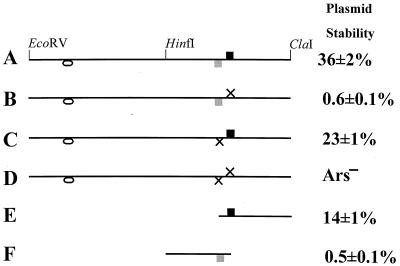
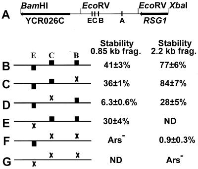
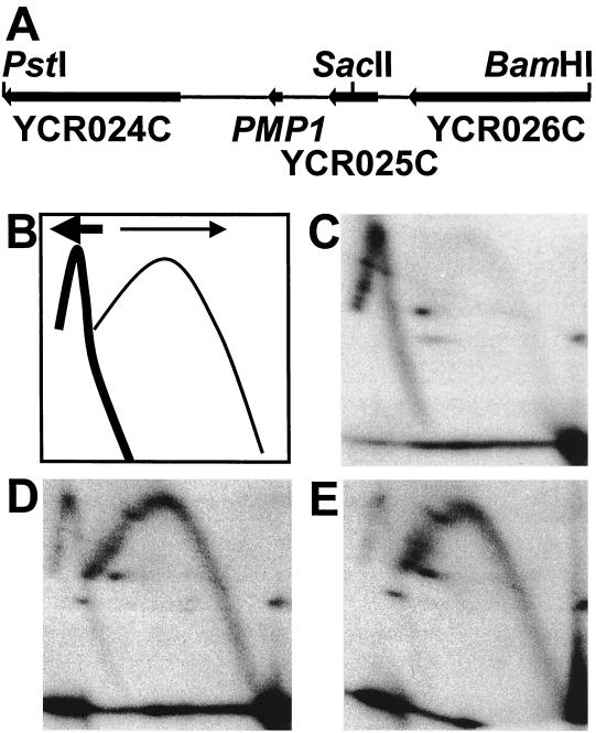
While many of the proteins involved in the initiation of DNA replication are conserved between yeasts and metazoans, the structure of the replication origins themselves has appeared to be different. As typified by ARS1, replication origins in Saccharomyces cerevisiae are <150 bp long and have a simple modular structure, consisting of a single binding site for the origin recognition complex, the replication initiator protein, and one or more accessory sequences. DNA replication initiates from a discrete site. While the important sequences are currently less well defined, metazoan origins appear to be different. These origins are large and appear to be composed of multiple, redundant elements, and replication initiates throughout zones as large as 55 kb. In this report, we characterize two S. cerevisiae replication origins, ARS101 and ARS310, which differ from the paradigm. These origins contain multiple, redundant binding sites for the origin recognition complex. Each binding site must be altered to abolish origin function, while the alteration of a single binding site is sufficient to inactivate ARS1. This redundant structure may be similar to that seen in metazoan origins.


Figures

FIG. 1
FIG. 1
Two ACS matches in ARS101. Line diagrams of ARS101 and mutant derivatives. The black box indicates the 11 of 11 match to the ACS (strong ORC binding site), and the grey box indicates the 9 of 11 match to the ACS (weak ORC binding site). The oval indicates an additional weak ORC binding site which does not contribute to ARS activity. X's indicate ACS knockout mutations. Plasmid stabilities are expressed as the percentage of plasmid-bearing cells present in a log-phase culture grown under selection. (A) WT 650-bp EcoRV-ClaI fragment. (B) The 11 of 11 match (Xba) mutant. (C) The 9 of 11 match (Msc) mutant. (D) Double mutant. (E) MscI-ClaI subclone, derived from C. (F) XbaI-HinFI subclone, derived from B.
FIG. 2
FIG. 2
2D gel analysis of chromosomal replication origin activity of ARS101 and mutant derivatives. (A) WT. (B) The 11 of 11 match mutant. (C) The 9 of 11 match mutant. (D) Double mutant. Arrows point to double-Y-shaped replication intermediates, indicative of termination. The inset in panel A is a schematic of replication intermediates as displayed by 2D gel analysis. The arc labeled B corresponds to bubble-shaped molecules, and the one labeled Y corresponds to Y-shaped molecules; the grey triangle labeled X corresponds to the region containing double-Y-shaped (termination) intermediates. (E) Diagram of the 5.3-kb EcoRI fragment examined by 2-D analysis. The positions of the SEN34 ORF and the included part of the adjacent Ty element are shown. The position of the 650-bp EcoRV-ClaI fragment is indicated by the grey box.
FIG. 3
FIG. 3
In vitro ORC footprint of ARS101. The left panel shows the footprint of WT ARS101 labeled near the ClaI end (see Fig. 1A). The black arrow marks the position of the 11 of 11 match to the ACS, and the grey arrow marks that of the 9 of 11 match. The region of protection resulting from ORC binding is indicated by the bracket, and the solid black arrowheads mark the positions of two relatively unaffected sites within this region. Lanes labeled 0 contain no ORC protein, and the triangle indicates lanes with increasing amounts of ORC (12.5, 25, 50, and 100 ng for the WT, 100 and 200 ng for the Xba mutant). R and T, A+G and T sequencing lanes, respectively. The black box indicates a weak ORC binding site near the top of the gel. The right panel shows the footprint of the 11 of 11 match mutant. Notice the lack of protection over that match. Protection (region delineated by the bracket) is seen over the 9 of 11 match (grey arrow), and the open arrowheads mark the positions of three hypersensitive sites induced by ORC binding to the 9 of 11 match.
FIG. 4
FIG. 4
Fork direction analyses flanking ARS101. (A) The upper line depicts the 13.9-kb BamHI fragment containing ARS101. The positions of the SEN34 ORF and YARCTy1-1 are indicated by arrows. The position of the 650-bp EcoRV-ClaI fragment containing ARS101 is shown by the grey box below the line. Below this are the 5.6-kb BamHI-BglII fragment, used to examine fork direction to the left of ARS101, and the 4.5-kb BglII-BamHI fragment, used on the right. The arrows mark the positions of the EcoRV and EcoRI sites used for in-gel digestion prior to the second dimension. The probes used are indicated below these lines. As discussed by Friedman and Brewer (22), in-gel digestion allows one to distinguish rightward-moving replication forks from leftward-moving ones. How these replication intermediates are resolved depends on the geometry of the origin, the site of digestion, and the probe used to detect them. (B) Schematic diagram for fork direction analysis to the left of ARS101. Replication intermediates from leftward-moving (thick arrow) forks, including those emanating from ARS101, are shown as a thick line. Intermediates from rightward-moving (thin arrow) forks, i.e., those moving towards ARS101 are shown as a thin line. (C) Schematic diagram for fork direction analysis to the right of ARS101. Replication intermediates from rightward-moving (thick arrow) forks, including those emanating from ARS101, are shown as a thick line. Intermediates from leftward-moving (thin arrow) forks, those moving towards ARS101, are shown as a thin line. (D) Left side, WT. (E) Right side, WT. (F) Left side, double mutant. (G) Right side, double mutant.
FIG. 5
FIG. 5
Three ACS matches contribute to ARS310 activity. (A) Diagram of the 2.2-kb BamHI-XbaI fragment containing ARS310. Arrows indicate the included portions of the YCR026C and RSG1 ORFs. The positions of ACS matches E, C, B, and A are marked by lines. The EcoRV sites defining the 0.85-kb fragment are indicated. (B to G) Plasmid constructs carrying the WT ARS310 and various ACS mutant derivatives. Black boxes represent the ACS matches, while X's denote knockout mutations; match A is not contained in the region shown and is unaltered in all the constructs. ND, not determined. Plasmid stabilities are reported for two contexts, the 0.85-kb EcoRV fragment (frag.) and the 2.2-kb BamHI-XbaI fragment. The larger fragment contains a stimulator of ARS activity, as indicated by the increased stabilities of all the constructs in the BamHI-XbaI context. While the BC double knockout appears to be Ars in the context of the EcoRV fragment, it is weakly active in the larger context (F). The triple mutant is Ars in this context (G).
FIG. 6
FIG. 6
Chromosomal replication origin activity of ARS310 and its mutant derivatives. (A) Diagram of the 5.3-kb HindIII fragment examined by 2D gels. The positions of the RSG1 and the included portion of the YCR026C ORFs are indicated by arrows. The position of a Gln tRNA gene is indicated by the arrowhead; this tRNA gene is in the correct position and orientation to cause the pause site which appears when ARS310 is inactivated. The grey box marks the position of the 0.85-kb EcoRV fragment containing ARS310. The lines within this box indicate the positions of the three matches to the ACS, B, C, and E. (B) WT. (C) B mutant. (D) C mutant. (E) BC double mutant. (F) BCE triple mutant. Arrows in B to F point to a spot resulting from the accumulation of Y-shaped replication intermediates, indicative of a pause site.
FIG. 6
FIG. 6
Chromosomal replication origin activity of ARS310 and its mutant derivatives. (A) Diagram of the 5.3-kb HindIII fragment examined by 2D gels. The positions of the RSG1 and the included portion of the YCR026C ORFs are indicated by arrows. The position of a Gln tRNA gene is indicated by the arrowhead; this tRNA gene is in the correct position and orientation to cause the pause site which appears when ARS310 is inactivated. The grey box marks the position of the 0.85-kb EcoRV fragment containing ARS310. The lines within this box indicate the positions of the three matches to the ACS, B, C, and E. (B) WT. (C) B mutant. (D) C mutant. (E) BC double mutant. (F) BCE triple mutant. Arrows in B to F point to a spot resulting from the accumulation of Y-shaped replication intermediates, indicative of a pause site.
FIG. 7
FIG. 7
In vitro ORC footprint of ARS310. The footprints of WT ARS310 and four mutant derivatives, single knockouts of ACS matches B and C, the BC double mutant, and the BCE triple knockout mutant, are shown. The positions of ACS matches B, C, and E are marked by the arrows on the right side. The uppermost brackets mark the region where ORC binding to match C versus match E is readily distinguished. The middle and lower brackets mark the regions protected by ORC binding to matches C and B, respectively. Hypersensitive sites are marked by dots. Labels on individual lanes are as in Fig. 3. The amounts of ORC used for the WT fragment were 10, 20, 40, and 60 ng, and for the mutant fragments they were 20 and 60 ng.
FIG. 8
FIG. 8
Fork direction analysis to the left of ARS310. (A) The 4.8-kb PstI-BamHI fragment examined in this analysis is shown. It is directly adjacent to the left end of the 2.2-kb BamHI-XbaI fragment shown in Fig. 5A. The positions of the YCR025C and PMP1 ORFs and the included portions of the YCR024C and YCR026C ORFs are shown. The SacII site used for in-gel digestion is also indicated. The 2.9-kb PstI-SacII fragment was used as a probe. (B) Schematic diagram for fork direction analysis to the left of ARS310. Replication intermediates arising from leftward-moving (thick arrow) forks, including those emanating from ARS310, are shown as a thick line. Intermediates from rightward-moving (thin arrow) forks, those moving towards ARS310, are shown as a thin line. (C) WT. (D) BC double mutant. (E) BCE triple mutant.

References

    1. Ausubel F M, Brent R, Kingston R E, Moore D D, Seidman J G, Smith J A, Struhl K, editors. Current protocols in molecular biology. New York, N.Y: John Wiley & Sons, Inc.; 1994.
    1. Becker D M, Guarente L. High-efficiency transformation of yeast by electroporation. Methods Enzymol. 1991;194:182–187. - PubMed
    1. Bell S P, Stillman B. ATP-dependent recognition of eukaryotic origins of DNA replication by a multiprotein complex. Nature. 1992;357:128–134. - PubMed
    1. Bielinsky A K, Gerbi S A. Chromosomal ARS1 has a single leading strand start site. Mol Cell. 1999;3:477–486. - PubMed
    1. Bielinsky A K, Gerbi S A. Discrete start sites for DNA synthesis in the yeast ARS1 origin. Science. 1998;279:95–98. - PubMed

Publication types