. 2001 Apr;21(8):2815-25.
doi: 10.1128/MCB.21.8.2815-2825.2001.

Reduction of target gene expression by a modified U1 snRNA

Affiliations

Reduction of target gene expression by a modified U1 snRNA

S A Beckley et al. Mol Cell Biol. 2001 Apr.

Abstract

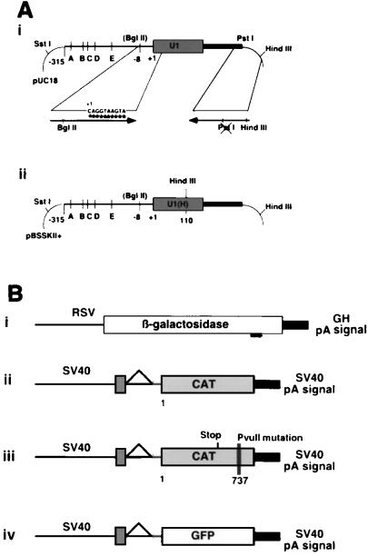
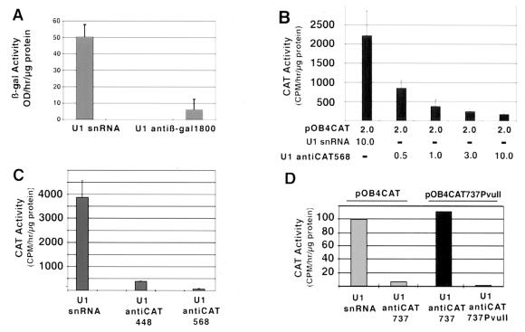
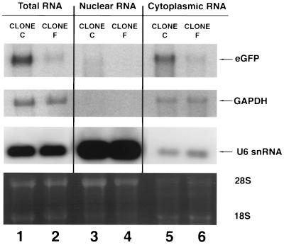
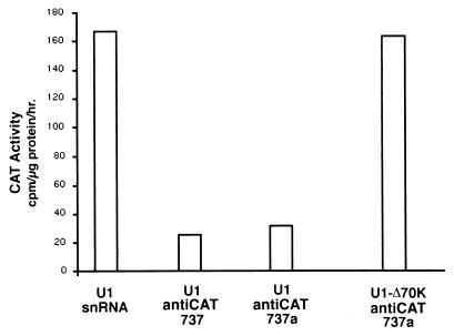
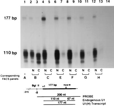
Although the primary function of U1 snRNA is to define the 5' donor site of an intron, it can also block the accumulation of a specific RNA transcript when it binds to a donor sequence within its terminal exon. This work was initiated to investigate if this property of U1 snRNA could be exploited as an effective method for inactivating any target gene. The initial 10-bp segment of U1 snRNA, which is complementary to the 5' donor sequence, was modified to recognize various target mRNAs (chloramphenicol acetyltransferase [CAT], beta-galactosidase, or green fluorescent protein [GFP]). Transient cotransfection of reporter genes and appropriate U1 antitarget vectors resulted in >90% reduction of transgene expression. Numerous sites within the CAT transcript were suitable for targeting. The inhibitory effect of the U1 antitarget vector is directly related to the hybrid formed between the U1 vector and target transcripts and is dependent on an intact 70,000-molecular-weight binding domain within the U1 gene. The effect is long lasting when the target (CAT or GFP) and U1 antitarget construct are inserted into fibroblasts by stable transfection. Clonal cell lines derived from stable transfection with a pOB4GFP target construct and subsequently stably transfected with the U1 anti-GFP construct were selected. The degree to which GFP fluorescence was inhibited by U1 anti-GFP in the various clonal cell lines was assessed by fluorescence-activated cell sorter analysis. RNA analysis demonstrated reduction of the GFP mRNA in the nuclear and cytoplasmic compartment and proper 3' cleavage of the GFP residual transcript. An RNase protection strategy demonstrated that the transfected U1 antitarget RNA level varied between 1 to 8% of the endogenous U1 snRNA level. U1 antitarget vectors were demonstrated to have potential as effective inhibitors of gene expression in intact cells.


Figures

FIG. 1
FIG. 1
(A) U1 snRNA locus. (i) Parental U1 snRNA construct with enhancer elements A through E. (ii) Map of the U1(H) construct. The arrows show the specific PCR primers used to introduce the mutations. (B) Target expression vectors. The pOB4 family of constructs has a single splice unit in which a cassette containing a triple stop unit (vertical lines) and the reporter gene are included in the terminal exon. (i) RSV β-Gal is a single exon construct; (ii) pOB4CAT; (iii) pOB4CAT(PvuII 737); (iv) pOB4GFP.
FIG. 2
FIG. 2
(A) Reduction in β-Gal activity from three separate transient U1 anti-β-Gal–RSV β-Gal cotransfection experiments. (B) Titration of various amounts of pOB4CAT to 10 μg of U1 anti-CAT568, yielding approximate molar ratio of CAT to U1 anti-CAT of 4:1, 2:1, 2:3, and 1:5 respectively. (C) Reduction of CAT enzyme activity by U1 anti-CAT448 and anti-CAT568 using cotransfection. (D) Sequence-specific reduction of CAT activity in transient U1 anti-CAT-pOB4CAT cotransfection experiments. pOB4CAT was used in lanes 1 and 2, and pOB4CAT 737PvuII was used in lanes 3 and 4. The U1 snRNA constructs were as follows: lane 1, U1 snRNA; lane 2, U1 anti-CAT737; lane 3, U1 anti-CAT737; lane 4, U1 anti-CAT737PvuII. These constructs were transiently cotransfected with TK-luciferase construct into NIH 3T3 cells. Error bars, standard deviations.
FIG. 3
FIG. 3
(A) Reduction of CAT activity from clonally expanded stable U1 anti-CAT568/pOB4CAT cotransfection experiments. Bars 1 to 6 represent the U1 snRNA-transfected CAT-expressing cells. Bars 7 to 12 represent the U1 anti-CAT transfected CAT-expressing cells. (B) CAT activity from a clonal cell line derived by stable pOB4CAT and subsequently transfected with U1 antiCAT568. Bars 1 to 5 represent the U1 snRNA-transfected clones, and bars 6 to 10 represent the U1 anti-CAT-transfected clones.
FIG. 4
FIG. 4
(A) Micrographs showing the reduction of stable GFP expression in NIH 3T3 cells by U1(H) anti-GFP. U1 Panels 1 and 3, U1 snRNA; panels 2 and 4, U1 anti-GFP. (B) FACS analysis of the cells shown in panel A shows the reduction in the number and intensity of the fluorescent cells containing the U1 anti-GFP construct. Panel 1, untransfected NIH 3T3 cells; panels 2 to 4, untransfected pOB4GFP stable line; panel 3, pOB4GFP and U1 snRNA; panel 4, pOB4GFP and U1 anti-GFP. (C) FACS of clonal lines stably transfected with control or U1 anti-GFP constructs. Panels: A, untransfected NIH 3T3 cells; B to H, pOB4GFP-expressing cells either untransfected (B) or transfected with U1(H) snRNA (C and D) or U1(H) anti-GFP (E to H).
FIG. 5
FIG. 5
Total RNA from an expanded clonal population of GFP-expressing cells stably transfected with U1(H) snRNA and U1(H) anti-GFP derived from the experiment shown in Fig. 4C. Clone C, U1(H) snRNA; clone F, U1(H) anti-GFP. GFP mRNA was normalized to GAPDH. U6 snRNA was used to show the efficiency of nuclear and cytoplasmic RNA extraction. The lower panel shows the ethidium bromide-stained agarose gel.
FIG. 6
FIG. 6
RNase protection assay showing the relative amounts of GFP transgenes in nuclear and cytoplasmic mRNA from clonal populations of stably GFP-expressing cells subsequently transfected with U1(H) snRNA or U1(H) anti-GFP. The properly terminated transgene is seen as a 350-nt band, and read-through mRNA is seen as a 500-bp protected band. The two controls are in lane 1 (total NIH 3T3 RNA) and lane 14 (tRNA). Lanes 2, 4, 6, 8, 10, and 12 contain nuclear RNA, and lanes 3, 5, 7, 9, 11, and 13 control cytoplasmic RNA. The following clones are those described in the FACS analysis in Fig. 4C: clone B, GFP-expressing cells (lanes 2 and 3); clone C, U1(H) snRNA (lanes 4 and 5); and clones E to H, U1(H) anti-GFP (in lanes 6 and 7, 8 and 9, 10 and 11, and 12 and 13, respectively).
FIG. 7
FIG. 7
Loss of inhibitory activity of the U1 anti-CAT737 construct when the 70K binding domain is destroyed. The constructs used to study the role of the 70K binding protein and PAP in vitro (30) were adapted to express the modified U1 transcripts from the U1 promoter. The control for an intact 70K binding domain (U1 anti-CAT737a) is identical to the U1 antiCAT737 analyzed in Fig. 2. These constructs were used in transient-cotransfection experiments with pOB4CAT and TK-luciferase in NIH 3T3 cells.
FIG. 8
FIG. 8
RNase protection assay showing the relative amount of U1 antitarget RNA in the nuclear (N) and cytoplasmic (C) mRNA from clonal populations of stably GFP-expressing cells subsequently transfected with U1 snRNA or U1(H) anti-GFP (Fig. 4C). The U1(H) snRNA transcript is a 177-nt band, and the endogenous U1 snRNA transcript is a 110-nt band. The two controls are in lane 1 (total NIH 3T3 RNA) and lane 14 (tRNA). Lanes 2, 4, 6, 8, 10, and 12 contain nuclear RNA, and lanes 3, 5, 7, 9, 11, and 13 contain cytoplasmic RNA. The following clones are those described in the FACS analysis in Fig. 4C: clone B, GFP-expressing cells (lanes 2 and 3); clone C, U1(H) snRNA (lanes 4 and 5); clones E to H, U1(H) anti-GFP (in lanes 6 and 7, 8 and 9, 10 and 11, and 12 and 13, respectively). The appearance of a U1 transcript in lane 3 (clone B cytoplasm) probably represents contamination of the sample with nuclear RNA, as suggested by its smaller size.

References

    1. Ashe M P, Furger A, Proudfoot N J. Stem-loop 1 of the U1 snRNP plays a critical role in the suppression of HIV-1 polyadenylation. RNA. 2000;6:170–177. - PMC - PubMed
    1. Ashe M P, Griffin P, James W, Proudfoot N J. Poly(A) site selection in the HIV-1 provirus: inhibition of promoter-proximal polyadenylation by the downstream major splice donor site. Genes Dev. 1995;9:3008–3025. - PubMed
    1. Ashe M P, Pearson L H, Proudfoot N J. The HIV-1 5′ LTR poly(A) site is inactivated by U1 snRNP interaction with the downstream major splice donor site. EMBO J. 1997;16:5752–5763. - PMC - PubMed
    1. Baker C C. An improved chloramphenicol acetyltransferase expression vector system for mapping transcriptional and post-transcriptional regulatory elements in animal cells. In: Alitalo K K, et al., editors. Recombinant systems in protein expression. Amsterdam, The Netherlands: Elsevier Science Publishers B. V.; 1990. pp. 75–86.
    1. Barksdale S K, Baker C C. The human immunodeficiency virus type 1 Rev protein and the Rev-responsive element counteract the effect of an inhibitory 5′ splice site in a 3′ untranslated region. Mol Cell Biol. 1995;15:2962–2971. - PMC - PubMed

Publication types

LinkOut - more resources