. 2001 Apr;21(8):2906-17.
doi: 10.1128/MCB.21.8.2906-2917.2001.

Discoidin domain receptor 1 tyrosine kinase has an essential role in mammary gland development

Affiliations

Discoidin domain receptor 1 tyrosine kinase has an essential role in mammary gland development

W F Vogel et al. Mol Cell Biol. 2001 Apr.

Abstract

Various types of collagen have been identified as potential ligands for the two mammalian discoidin domain receptor tyrosine kinases, DDR1 and DDR2. Here, we used a recombinant fusion protein between the extracellular domain of DDR1 and alkaline phosphatase to detect specific receptor binding sites during mouse development. Major sites of DDR1-binding activity, indicative of ligand expression, were found in skeletal bones, the skin, and the urogenital tract. Ligand expression in the uterus during implantation and in the mammary gland during pregnancy colocalized with the expression of the DDR1 receptor. The generation of DDR1-null mice by gene targeting yielded homozygous mutant animals that were viable but smaller in size than control littermates. The majority of mutant females were unable to bear offspring due to a lack of proper blastocyst implantation into the uterine wall. When implantation did occur, the mutant females were unable to lactate. Histological analysis showed that the alveolar epithelium failed to secrete milk proteins into the lumen of the mammary gland. The lactational defect appears to be caused by hyperproliferation and abnormal branching of mammary ducts. These results suggest that DDR1 is a key mediator of the stromal-epithelial interaction during ductal morphogenesis in the mammary gland.


Figures

FIG. 1
FIG. 1
Expression of DDR1-binding activity, shown by detection of DDR1-binding sites using a DDR1-hAP fusion protein. (A) Staining of a transverse section of a 6-day-old mouse with DDR1-hAP. DDR1-binding sites were detected in the skeleton, skin, and urogenital tract. (B to D) Staining of a molar tooth (B), the whisker barrel (C), and the periosteal collar of the clavicle (D). (E to G) Staining of a parasagittal section of an E13.5 mouse embryo with DDR1-hAP. Strong signals are seen in the ribs (E), the cartilage primordium of the metatarsal bones (F), and at higher magnification in the periosteum of the ribs (G). (H to K) Colocalization of DDR1-binding affinity and collagen expression in the mouse mammary gland. (H) Staining of mammary epithelial cells and adjacent myofibroblasts with DDR1-hAP using sections from a pregnant female at day 12.5 of gestation. (J) The binding of DDR1-hAP is competed with recombinant extracellular domain of DDR1. (K) Immunostaining for collagen type III in the mammary gland (brownish color).
FIG. 2
FIG. 2
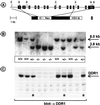
Generation of DDR1-null mice. (A) Organization of the mouse DDR1 gene (top) and the targeting construct to generate DDR1-null mice (bottom). Exons are indicated by black boxes that are numbered beginning with the first coding exon as exon 1. The lengths of exons and introns are not to scale. Relevant restriction sites are indicated: E, EcoRI; B, BamHI; X, XhoI; N, NotI. The checkered box represents the external probe used for genotyping of ES cells and mice. (B) Southern blot analysis of DNA isolated from the embryos of a heterozygous intercross (+/+, wild-type; +/−, heterozygous; −/−, homozygous). An 8.0-kb BamHI fragment corresponds to the wild-type allele, and a 3.8-kb fragment corresponds to the mutant allele. (C) Western blot analysis of lysates generated from the same embryos as in panel B. DDR1 is detected as an approximately 130-kDa protein. Asterisks denote heterozygous embryos with reduced DDR1 expression.
FIG. 3
FIG. 3
Dwarfism of DDR1-null mice. (A) Growth curves of male and female offspring during the first 10 weeks of age. One set of growth curves, which is representative of six measurements taken from different litters during the same time period, is depicted. (B and C) X-ray analysis of heterozygous (B) and mutant (C) 10-week-old females. The lack of proper mineralization of the fibula bone is shown by an arrow in the insert. (D and E) Hematoxylin-fast green-safranin O staining of growth plates in heterozygous (D) and mutant (E) metatarsal bones of 2-week-old mice. The comparable length of the growth plate in the mutant and control section is indicated by an arrow. (F and G) BrdU-stained sections of tibias from 2-week-old heterozygous (F) and mutant (G) mice. Arrows mark the length of the proliferative zone. (H to N) TUNEL assay with sections from the tibia of 4-week-old wild-type (H, K, M) and mutant (J, L, N) animals. No increase in chondrocyte apoptosis was seen in DDR1-null chondrocytes (H and J). Sections were pretreated with DNase I as positive control (K and L) or incubated without enzyme as negative control (M and N).
FIG. 4
FIG. 4
Defects of DDR1-null mice in ear and placental development. (A) Phenotypic appearance of ears in control and mutant mice. Mice were 3 months of age and were anesthetized prior to photography. (B) Detection of DDR1-binding affinity in the ear cartilage using the DDR1-hAP fusion protein (indicated by arrows). The hair follicles are stained as well. (C to F) DDR1-hAP staining (C and E) and DDR1 immunostaining (D and F) in pre- (E2.5; C and D) and post- (E4.5; E and F) implantational uteri in wild-type mice. Abbreviations: mm, myometrium; lm, longitudinal muscle; st, stroma; gl, glandular epithelium; dz, desidual zone; em, embryo proper.
FIG. 5
FIG. 5
Mammary gland defects in DDR1-null mice. (A and B) Hematoxylin-eosin stain of late pregnancy (day 18.5 of gestation) mammary glands of heterozygous (A) and mutant (B) mice. (C to F) Whole-mount analysis of control (C and E) and mutant (D and F) mammary glands of day 18.5 pregnant females. Pictures are taken at low (C and D) and higher (E and F) magnification. (G and H) Mammary gland morphology of heterozygous (G) and mutant (H) mice 1 day postpartum. Note the almost complete absence of milk secretion into the lumens in panel H. (J and K) Regressed mammary epithelium in mutant mice (K) compared to that in control mice (J) 2 days postpartum.
FIG. 6
FIG. 6
Mammary gland development during puberty and pregnancy in DDR1-null mice. (A and B) Whole-mount analysis of the developing mammary gland in 3-week-old heterozygous (A) and mutant (B) mice. Some terminal end buds are marked by arrows. (C and D) Masson-Goldner stain of virgin heterozygous (C) and mutant (D) mammary glands. Note the enlarged ducts and the increase in extracellular matrix deposition in the adipose tissue (insert). (E and F) Ki-67 staining of virgin control (E) and mutant (F) mammary gland. The number of Ki-67-positive (red-labeled) nuclei is approximately five times higher in the mutant tissue. (G) Immunolocalization of DDR1 in a lactating mammary gland. Specific staining (brownish color) is seen in the luminal epithelial cells. (H) Western blot analysis of DDR1 expression during pregnancy and lactation.
FIG. 7
FIG. 7
Expression of milk proteins in DDR1-null females. (A) Western blot analysis of milk protein expression in lysates from mouse mammary glands 2 days postpartum. Arrows indicate milk proteins reduced or absent in DDR1-null animals. Northern blot analysis of β-casein (B) and α-lactalbumin together with WDNM1 (C) expression during mammary gland development of mutant and control mice. The lower strip in panel B shows the ethidium bromide-stained gel, and the lower strip in panel C shows reanalysis of the blot with a 28S rRNA probe.

Similar articles

Cited by

References

    1. Alves F, Vogel W, Mossie K, Millauer B, Höfler H, Ullrich A. Distinct structural characteristics of discoidin I subfamily receptor tyrosine kinases and complementary expression in human cancer. Oncogene. 1995;10:609–618. - PubMed
    1. Aszódi A, Chan D, Hunziker E, Bateman J F, Fässler R. Collagen II is essential for the removal of the notochord and the formation of invertebral discs. J Cell Biol. 1998;30:1399–1412. - PMC - PubMed
    1. Barker K T, Martindale J E, Mitchell P J, Kamalati T, Page M J, Phippard D J, Dale T C, Guterson B A, Crompton M R. Expression patterns of the novel receptor-like tyrosine kinase, DDR, in human breast tumors. Oncogene. 1995;10:569–575. - PubMed
    1. Baumgartner S, Hofmann K, Chiquet-Ehrismann R, Bucher P. The discoidin domain family revisited: new members from prokaryotes and homology-based fold prediction. Protein Sci. 1998;7:1626–1631. - PMC - PubMed
    1. Bhatt R S, Tomoda T, Fang Y, Hatten M E. Discoidin domain receptor 1 functions in axon extension of cerebellar granule neurons. Genes Dev. 2000;14:2216–2228. - PMC - PubMed

Publication types

MeSH terms