. 2001 Apr;21(8):2956-66.
doi: 10.1128/MCB.21.8.2956-2966.2001.

Cyclin A is a mediator of p120E4F-dependent cell cycle arrest in G1

Affiliations

Cyclin A is a mediator of p120E4F-dependent cell cycle arrest in G1

L Fajas et al. Mol Cell Biol. 2001 Apr.

Abstract

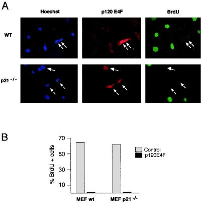
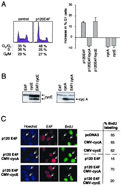
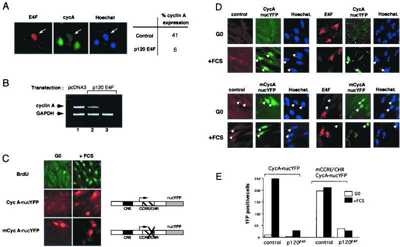
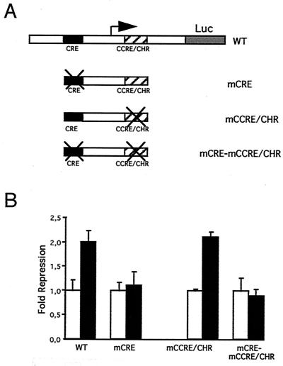
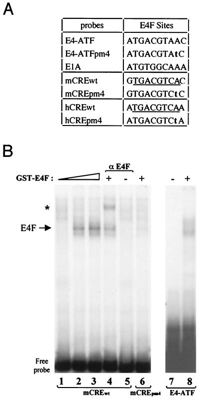
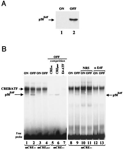
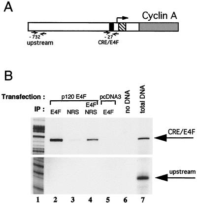
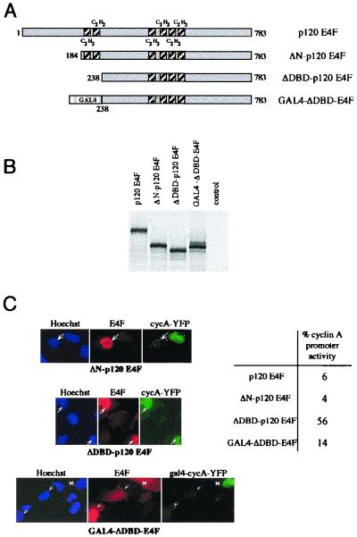
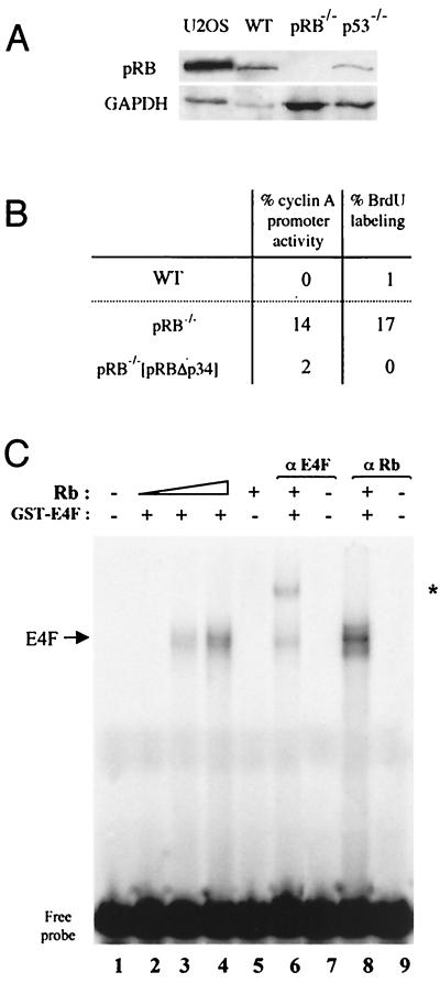
E4F is a ubiquitously expressed GLI-Krüppel-related transcription factor which has been identified for its capacity to regulate transcription of the adenovirus E4 gene in response to E1A. However, cellular genes regulated by E4F are still unknown. Some of these genes are likely to be involved in cell cycle progression since ectopic p120E4F expression induces cell cycle arrest in G1. Although p21WAF1 stabilization was proposed to mediate E4F-dependent cell cycle arrest, we found that p120E4F can induce a G1 block in p21(-/-) cells, suggesting that other proteins are essential for the p120E4F-dependent block in G1. We show here that cyclin A promoter activity can be repressed by p120E4F and that this repression correlates with p120E4F binding to the cyclic AMP-responsive element site of the cyclin A promoter. In addition, enforced expression of cyclin A releases p120E4F-arrested cells from the G1 block. These data identify the cyclin A gene as a cellular target for p120E4F and suggest a mechanism for p120E4F-dependent cell cycle regulation.


Figures

FIG. 1
FIG. 1
p120E4F -dependent cell cycle arrest is maintained in p21WAF1−/− MEFs. WT or p21WAF1−/− MEFs were transfected with a p120E4F expression vector and BrdU labeled. The panels show Hoechst staining, E4F immunodetection in E4F-transfected cells, and BrdU staining of cells undergoing DNA synthesis (A). Quantitation of the BrdU-labeled cells that either overexpressed p120E4F (p120E4F) or did not (control) is schematized for both WT and p21WAF1−/− MEFs (B).
FIG. 2
FIG. 2
Enforced expression of cyclin A, but not of cyclin E, releases cells from the G1 block induced by overexpressed p120E4F. (A) p120E4F induces a G1 arrest which is released by coexpression of cyclin A. (Left) U2OS cells were transfected with plasmids expressing p120E4F (10 μg) and the CD20 marker (1μg). Cells were harvested 36 h after transfection, and DNA profiles of CD20-positive cells were obtained using bivariate flow cytometry. Percentage of cells present in the different cell cycle phases is indicated below each plot. (Right) U2OS cells were transfected with plasmids expressing p120E4F and the CD20 marker alone or in combination with a cyclin A (0.1 μg) or cyclin E (0.1 μg) expression vector. As a control, U2OS cells were also transfected with CD20 expression plasmid in combination with either cyclin A or cyclin E expression plasmid. Absolute increase in percentage of U2OS cells in the G0/G1 phase upon expression of the indicated proteins is plotted on the y axis, the baseline representing the percentage of G0/G1 cells in mock-transfected cells. Data are representative of at least three independent experiments. (B) Total cellular extracts from CD20-positive cells were submitted to Western blot analysis using an antibody directed at cyclin E (left) or cyclin A (right). Arrows point to specific bands. (C) U2OS cells overexpressing both p120E4F and cyclin A resume DNA synthesis. U2OS cells were cotransfected with plasmids expressing p120E4F in combination with either cyclin A or cyclin E. The panels show Hoechst staining of nuclei, E4F immunodetection in E4F-transfected cells (indicated by arrows), and BrdU staining of cells undergoing DNA synthesis. The percentage of BrdU-labeled cells (85 counted cells) is indicated for cells expressing p120E4F alone or in combination with either cyclin A (p120E4F/CMV-cycA) or cyclin E (p120E4F/CMV-cycE). As a control, the index of BrdU labeling was measured for cells transfected with either cyclin A or cyclin E expression plasmids.
FIG. 3
FIG. 3
p120E4F represses expression of cyclin A. (A) p120E4F overexpression decreases cyclin A cellular concentrations. U2OS cells were transfected with p120E4F expression plasmid and immunostained for overexpressed p120E4F and endogenous cyclin A. The panels show immunodetection of E4F and endogenous cyclin A as well as Hoechst staining. The percentage of cyclin A-expressing cells, depending on whether they overexpress p120E4F (p120E4F) or not (control), is indicated (50 cells counted). (B) p120E4F overexpression decreases cyclin A mRNA concentrations. NIH 3T3 cells were transfected with p120E4F expression plasmid (lanes 2 and 3) or with pcDNA3 control vector (lane 1). mRNA was amplified by RT-PCR using, in the same PCR, primers to amplify both the cyclin A and the control GAPDH loci (lanes 1 and 2). In lane 3, cyclin A primers were omitted from the PCR in order to further confirm the identification of the cylin A amplified DNA fragment. (C) The YFP gene fused to the cyclin A promoter recapitulates cyclin A transcriptional regulation during the cell cycle. Mutation of the CCRE/CHR module leads to expression of the reporter in both quiescent (G0) and stimulated (+FCS) cells. Plasmid pCycA-nucYFP, encoding YFP tagged with an NLS and placed under the transcriptional control of the murine cyclin A promoter, was used in transient transfections. After transfection, cells were split in half; each half was submitted to serum starvation (24 h), followed by either serum refeeding (16 h; +FCS) or further starvation (16 h; -FCS). In parallel, cells were transfected with the expression plasmid pmCycA-nucYFP, where the WT cyclin A promoter was replaced by a cyclin A promoter mutated at the CCRE/CHR site. The panels show BrdU staining as well as expression of YEP in the nuclei. (D) p120E4F overexpression represses both WT (upper panels) and mutated (lower panels) cyclin A promoters. CCL39 cells were cotransfected with p120E4F and either pCycA-nucYFP or pmCycA-nucYFP expression vector. The panels show E4F immunodetection, YFP expression, and Hoechst staining in E4F-transfected serum-starved (G0) or serum-restimulated (+FCS) cells. Arrows and asterisks point to YFP-and E4F-expressing cells, respectively. (E) Number of YFP-expressing cells, plotted for a representative experiment, in serum-starved (G0) or serum-restimulated (+FCS) cells. Cells were cotransfected with pCycA-nucYFP or pmCycA-nucYFP together with respectively an empty (control) or a p120E4F -expressing (p120E4F) vector.
FIG. 4
FIG. 4
p120E4F overexpression represses cyclin A transcription. (A) Luciferase reporter gene under the transcriptional regulation of the murine cyclin A promoter. The various cyclin A promoter constructs tested, which include the WT promoter as well as the promoter mutated at the CRE (mCRE), at the CCRE/CHR (mCCRE/CHR) or at both (mCRE-mCCRE/CHR), are schematized. (B) Luciferase activities of the various cyclin A promoter constructs were measured in CCL39 cells cotransfected with either a p120E4F-expressing vector (filled bars) or an empty vector (empty bars). The fold repression of cyclin A promoter activity in response to overexpression of p120E4F is shown. Standard deviations are indicated for experiments done in triplicate.
FIG. 5
FIG. 5
The CRE of the cyclin A promoter is a p120E4F binding site. (A) Sequences of E4F binding sites found in E4 and E1A promoters. The E4-ATFpm4 point mutation abolishes E4F binding to DNA. The CRE sites found in the murine (mCREwt) and human (hCREwt) cyclin A promoters are indicated. The corresponding pm4 mutations are indicated in bold characters; the consensus CRE is underlined. (B) Binding of the purified p120E4F protein to cyclin A CRE. Purified GST-p120E4F was incubated with either the WT (lanes 1 to 5) or pm4 mutant (lane 6) murine cyclin A CRE probe. As a control, GST-E4F binding was tested on the E4-ATF site (lanes 7 and 8). DNA-protein complexes were analyzed by EMSA. The E4F complex is indicated, as is the E4F-containing complex (∗) supershifted with anti-p120E4F antibodies.
FIG. 6
FIG. 6
E4F binds to the CRE-E4F site of the cyclin A promoter independently of CREB and ATF proteins. (A) Expression of p50E4F in tetracycline-inducible cell lines. Cultures of TTN5-p50E4F cells were grown for 24 h in the presence (ON) or absence (OFF) of tetracycline. The blotted membrane was probed with anti-E4F antibodies, and antibody-antigen complexes were visualized by enhanced chemiluminescence. The Western blot shows the specific expression of p50E4F upon removal of tetracycline (OFF), whereas basal levels of E4F in the inducible TTN5-p50E4F cell line in the presence of tetracycline (ON) were undetectable. (B) Evidence that E4F present in nuclear extracts can bind to the CRE-E4F site of the cyclin A promoter. Nuclear extracts (1 μg) from TTN5-p50E4F cells grown in the presence (ON) or absence (OFF) of tetracycline were incubated with mCREwt (lanes 1, 2, and 5 to 13) or mCREpm4 (lanes 5 to 7) and subjected to EMSA. Competition was done with a 500-fold molar excess of unlabeled oligonucleotides corresponding to the WT murine cyclin A CRE site (lane 5), to the pm4 mutant site (lane 6), or to the E4-ATF site previously characterized (lane 7). Gel shift reactions were done in the presence of 1 μl of NRS (lanes 10 and 11) or rabbit anti-E4F antibodies (αE4F; lanes 12 and 13). Positions of migration of the CREB-ATF and p50E4F DNA-protein complexes and of the free probe are indicated.
FIG. 7
FIG. 7
p120E4F binds to the CRE-E4F site of the cyclin A promoter in vivo. (A) Chromatin immunoprecipitation experiments were performed with NIH 3T3 cells that were transfected either with p120E4F expression plasmid or with the empty pcDNA3 vector. Immunoprecipitations were then carried out with either anti-E4F rabbit antibodies or NRS. Two loci of the murine cyclin A promoter were checked by PCR amplification of the immunoprecipitated chromatin. The CRE-E4F site (black box) is positioned 27 bp upstream of the most 3′ transcription initiation site (arrow); a presumably irrelevant locus was chosen 732 bp upstream of this same transcription initiation site (upstream) as a control for immunoprecipitation specificity. The CCRE/CHR site is represented by a hatched box, while cyclin A coding sequence is in grey. (B) PCR amplification products of E4F chromatin immunoprecipitations were analyzed on a 6% denaturing gel along with a sequence ladder. (Top) amplification at the CRE-E4F locus; (bottom) results obtained with primers at the upstream locus. PCRs in lanes 2, 3, 4, and 7 were obtained with chromatin DNA purified from E4F-transfected cells, while the reaction in lane 5 was obtained from DNA of mock-transfected NIH 3T3 cells. Antibodies used for the immunoprecipitations (IP) are indicated. In lane 4, the supernatant of the immunoprecipitation done with NRS (lane 3) was further immunoprecipitated with anti-E4F antibodies. The PCR in lane 7 (total DNA) was performed with an aliquot of the DNA obtained from E4F-transfected cells taken prior to immunoprecipitation. The sequence ladder is shown in lane 1.
FIG. 8
FIG. 8
p120E4F DNA binding activity is required for cyclin A transcriptional repression. (A) Amino-terminal deletion mutants of p120E4F. Deletion of the first 183 aa of p120E4F (ΔN-p120E4F mutant) preserves the integrity of the six zinc finger domains. The ΔDBD-p120E4F mutant corresponds to a polypeptide (aa 238 to 783) which lacks the two p120E4F amino-terminal zinc finger motifs. The C2H2 zinc finger motifs are shown as shaded boxes. The GAL4-ΔDBD-E4F fusion protein contains the GAL4 DNA binding domain (aa 1 to 147) fused N terminally to ΔDBD-p120E4F. (B) Expression of the mutants in vitro. Full-length p120E4F as well as the mutants ΔN-p120E4F, ΔDBD-p120E4F, and GAL4-ΔDBD-E4F were in vitro translated and analyzed by SDS-PAGE along with unprogrammed reticulocyte lysates (control). (C) The two p120E4F N-terminal zinc finger domains are required for repression of cyclin A transcription. U2OS cells were cotransfected with expression vectors for either pCycA-nucYFP and p120E4F, ΔN-p120E4F, or ΔDBD-p120E4F or for gal4-cycA-YFP and GAL4-ΔDBD-E4F. The panels show E4F immunodetection, YFP expression, and Hoechst staining. Arrows indicate E4F-transfected cells. A transfected cell with a cytosolic localization of GAL4-ΔDBD-E4F fusion protein is indicated by the asterisk. The percentage of cells that contain an active cyclin A promoter, as estimated by expression of YFP, is indicated (about 100 counted cells).
FIG. 9
FIG. 9
p120E4F overexpression represses cyclin A transcription independently of p53. (A) Absence of UV-induced p53 expression in p53−/− MEFs. Cells were submitted to UV irradiation (20 J/cm2) and further grown for 8 h before fixation. The panels show p53 immunostaining in WT or p53−/− MEFs. (B) p53−/− or WT MEFs were cotransfected with p120E4F and pCycA-nucYFP expression vectors. The panels show E4F immunodetection in E4F-transfected cells, YFP expression under the control of the murine cyclin A promoter, BrdU labeling, and Hoechst staining. (C) The percentage of mock- or E4F-transfected cells that contain an active cyclin A promoter as determined by YFP expression or that are BrdU labeled is indicated for both WT and p53−/− MEFs. Three independent transfection experiments were analyzed.
FIG. 10
FIG. 10
p120E4F -dependent transcriptional repression of the cyclin A gene is enhanced by pRB. (A) pRB Western blot of cellular extracts from U2OS or either WT, pRB−/−, or p53−/− MEFs. The blot was probed with anti-pRB antibodies and reprobed with anti-GAPDH antibodies for normalization. (B) Absence of pRB results in loss of efficiency for E4F-dependent repression of cyclin A transcription. pRB−/− or WT MEFs were cotransfected with p120E4F and pCycA-nucYFP expression vectors. The percentage of E4F-transfected cells that contain an active cyclin A promoter or that are BrdU labeled is indicated for WT and pRB−/− MEFs as well as for pRB−/− MEFs cotransfected with pRBΔp34 expression plasmid. (C) Binding of purified p120E4F protein to the cyclin A CRE site is enhanced by pRB. Purified GST-p120E4F was incubated with the WT murine cyclin A CRE probe and with increasing concentrations of purified baculovirus-expressed pRB protein (lanes 2 to 4). The DNA-protein complex obtained with both p120E4F and pRB (lane 4) was incubated with antibodies directed against either E4F (lane 6) or pRB (lane 8). DNA-protein complexes were analyzed by EMSA. The E4F-containing complex supershifted with anti-p120E4F antibodies is indicated (∗).

References

    1. Barlat I, Fesquet D, Brechot C, Henglein B, Dupuy d'Angeac A, Vie A, Blanchard J M. Loss of the G1-S control of cyclin A expression during tumoral progression of Chinese hamster lung fibroblasts. Cell Growth Differ. 1993;4:105–113. - PubMed
    1. Barlat I, Henglein B, Plet A, Lamb N, Fernandez A, McKenzie F, Pouyssegur J, Vie A, Blanchard J M. TGF-beta 1 and cAMP attenuate cyclin A gene transcription via a cAMP responsive element through independent pathways. Oncogene. 1995;11:1309–1318. - PubMed
    1. Brugarolas J, Chandrasekaran C, Gordon J I, Beach D, Jacks T, Hannon G J. Radiation-induced cell cycle arrest compromised by p21 deficiency. Nature. 1995;377:552–557. - PubMed
    1. Chambard J C, Pognonec P. A reliable way of obtaining stable inducible clones. Nucleic Acids Res. 1998;26:3443–3444. - PMC - PubMed
    1. Chin J W, Kohler J J, Schneider T L, Schepartz A. Protein escorts to the transcription ball. Curr Biol. 1999;9:R929–R932. - PubMed

Publication types

MeSH terms

LinkOut - more resources