. 2001 Apr;13(4):781-92.
doi: 10.1105/tpc.13.4.781.

Demonstration in yeast of the function of BP-80, a putative plant vacuolar sorting receptor

Affiliations

Demonstration in yeast of the function of BP-80, a putative plant vacuolar sorting receptor

D Humair et al. Plant Cell. 2001 Apr.

Abstract

BP-80, later renamed VSR(PS-1), is a putative receptor involved in sorting proteins such as proaleurain to the lytic vacuole, with its N-terminal domain recognizing the vacuolar sorting determinant. Although all VSR(PS-1) characteristics and in vitro binding properties described so far favored its receptor function, this function remained to be demonstrated. Here, we used green fluorescent protein (GFP) as a reporter in a yeast mutant strain defective for its own vacuolar receptor, Vps10p. By expressing VSR(PS-1) together with GFP fused to the vacuolar sorting determinant of petunia proaleurain, we were able to efficiently redirect the reporter to the yeast vacuole. VSR(PS-1) is ineffective on GFP either alone or when fused with another type of plant vacuolar sorting determinant from a chitinase. The plant VSR(PS-1) therefore interacts specifically with the proaleurain vacuolar sorting determinant in vivo, and this interaction leads to the transport of the reporter protein through the yeast secretory pathway to the vacuole. This finding demonstrates VSR(PS-1) receptor function but also emphasizes the differences in the spectrum of ligands between Vps10p and its plant equivalent.


Figures

Figure 1.
Figure 1.
Constructs Used in This Study. Sec-GFP, the secreted form of GFP in which the Arabidopsis chitinase B signal peptide (s.p.) was fused to GFP5 (GFP); CPY-GFP, the signal peptide and the N-terminal propeptide from the yeast CPY (detailed) were fused to GFP5; GFP-Chi, the Ct-VSD of the tobacco chitinase A (detailed) was fused to the secreted form of GFP (Sec-GFP); aleu-GFP, the CPY signal peptide was fused with petunia aleurain VSD (detailed) to the N terminus of GFP5. For comparison (in parentheses), the corresponding sequence of the previously described VSD from barley proaleurain (Holwerda et al., 1992) is shown. Underlined amino acids are essential for vacuolar targeting.
Figure 2.
Figure 2.
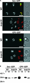
Vps10p-Dependent Vacuolar Localization of Sec-GFP and CPY-GFP in Yeast. (A) The living cells were visualized with a confocal microscope after 24 hr of induction. The constructs, either a secreted form of GFP (Sec-GFP) or a GFP fused to the yeast vacuolar signal from CPY (CPY-GFP), were expressed in wild-type cells (WT) or in a yeast disrupted for the gene encoding the vacuolar receptor Vps10p (Δvps10). Each condition combines four images from the same cells: from left to right, GFP fluorescence, FM 4-64 staining of a vacuolar membrane, a pseudocolored merged image with GFP in green and FM 4-64 in red, and the transmitted light image. formula image. (B) After 24 hr of induction, the transformed cells were separated in two fractions: the cell extract (c) and the medium (m). Both extracts then were immunolabeled with anti-GFP antibodies. Molecular masses of standards are indicated to the left in kilodaltons.
Figure 3.
Figure 3.
Demonstration of the Vacuolar Sorting Properties of the Petunia Aleurain Propeptide in Tobacco Leaf Protoplasts. Living protoplasts expressing aleuPh-GFP6 were visualized using a confocal microscope 24 hr after transformation. Two cells are shown after illumination with either the dual fluorescein isothiocyanate/tetramethylrhodamine isothiocyanate fluorescence filter (A) or transmitted light (B). The fluorescence image uses the attributed pseudocolors green for GFP and red for chloroplast autofluorescence. formula image.
Figure 4.
Figure 4.
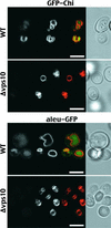
Vps10p-Dependent Vacuolar Localization of GFP-Chi and aleu-GFP in Yeast. The living cells were visualized with a confocal microscope after 24 hr of induction. The constructs, either a GFP fused to the tobacco chitinase A VSD (GFP-Chi) or the petunia aleurain VSD fused to GFP (aleu-GFP), were expressed in wild-type cells (WT) or a yeast disrupted for the gene encoding the vacuolar receptor Vps10p (Δvps10). Each condition combines four images from the same cells as described for Figure 2A. formula image.
Figure 5.
Figure 5.
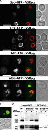
The Expression of VSRPS-1 Specifically Restores the Vacuolar Location of aleu-GFP in the Absence of the Yeast Vacuolar Sorting Receptor Vps10p. (A) The living cells were visualized with a confocal microscope after 24 hr of induction. The Δvps10 strain was doubly transformed with the plant putative vacuolar sorting receptor VSRPS-1 and one of the four GFP fusion constructs. These GFP chimera carry no vacuolar signal (Sec-GFP), the CPY yeast vacuolar signal (CPY-GFP), the tobacco chitinase A VSD (GFP-Chi), or the petunia aleurain VSD (aleu-GFP). Each condition combines four images from the same cells as described for Figure 2A. formula image. (B) Effect of VSRPS-1 on aleu-GFP localization in Δvps10 cells after 48 hr of induction. Shown are (top) a fluorescence image of a Δvps10 cell expressing both VSRPS-1 and aleu-GFP (green) and stained for the vacuolar membrane with FM 4-64 (red) and (bottom) the same cell in transmitted light. formula image. (C) Immunoblot analysis after 48 hr of induction by using anti-GFP antibodies on a cell extract from either wild-type yeast (WT) or a yeast disrupted for the gene encoding the vacuolar receptor Vps10p (Δvps10) and expressing the GFP reporter fused to either the petunia aleurain (aleu-GFP) or the chitinase A (GFP-Chi) VSD in the presence (+) or absence (−) of VSRPS-1. The top and bottom panels represent the same blots exposed for either 5 or 15 sec, respectively. Molecular masses of standards are indicated to the left in kilodaltons.
Figure 6.
Figure 6.
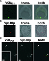
The Plant Vacuolar Sorting Receptor VSRPS-1 Is Not Vacuolar and Colocalizes with Vps10p in Yeast Cells. Cells were fixed after 24 hr of culture and immunolabeled with anti-VSR and/or anti-Vps10p antibodies. The resulting immunostaining was visualized using a confocal microscope. (A) A Δvps10 cell expressing VSRPS-1 was immunolabeled with anti-VSR antibodies (left). A transmitted image of the same cell (trans.) was taken and merged to the immunostaining image (both). (B) A wild-type cell was immunolabeled with anti-Vps10p antibodies (left). A transmitted image of the same cell (trans.) was taken and merged to the immunostaining image (both). (C) A wild-type cell expressing VSRPS-1 was immunolabeled with anti-VSR and anti-Vps10p antibodies. The immunolabeling images were pseudocolored in green for VSRPS-1 and red for Vps10p and merged (both). The inset shows the background staining with anti-VSR antibodies on untransformed wild-type cells. Arrowheads mark the location of double-labeled compartments. formula image.
Figure 7.
Figure 7.
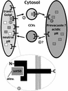
Model for VSRPS-1 Function in Plants. The trans-Golgi (left) is the region of the secretory pathway where sorting of vacuolar soluble proteins is believed to occur. At this neutral pH, the plant vacuolar sorting receptor (brackets) sorts (step 1) proaleurain (gray squares) from other soluble proteins (white squares). The detail shows more precisely the structure of VSRPS-1, with its N-terminal domain (N) interacting with the ssVSD carried by proaleurain (aleu.). It also shows the C-terminal domain (C) of VSRPS-1 on the cytosolic side of the membrane that is available for contacts with proteins such as adaptins that are involved in CCV formation. The complex of VSRPS-1 and proaleurain (step 2) is packed into transport vesicles and targeted to an acidic compartment (right). The decrease in pH causes the ligand to be released from VSRPS-1 (step 3) within the acidic compartment, which is possibly a prevacuole. The free receptor then recycles back to the Golgi (step 4), again by means of transport vesicles. We know that CCVs are used by the receptor in plants to shuttle between the Golgi and an acidic compartment, but we do not know in which direction of transport. This model is based on our demonstration of VSRPS-1 function in yeast and on previously published characteristics (Kirsch et al., 1994; Paris et al., 1997).

Similar articles

Cited by

References

    1. Abedi, M.R., Caponigro, G., and Kamb, A. (1998). Green fluorescent protein as a scaffold for intracellular presentation of peptides. Nucleic Acids Res. 26, 623–630. - PMC - PubMed
    1. Ahmed, S.U., Bar-Peled, M., and Raikhel, N.V. (1997). Cloning and subcellular location of an Arabidopsis receptor-like protein that shares common features with protein-sorting receptors of eukaryotic cells. Plant Physiol. 114, 325–336. - PMC - PubMed
    1. Bednarek, S., and Raikhel, N.V. (1991). The barley lectin carboxyl-terminal propeptide is a vacuolar protein sorting determinant in plants. Plant Cell 3, 1195–1206. - PMC - PubMed
    1. Blum, S., Mueller, M., Schmid, S.R., Linder, P., and Trachsel, H. (1989). Translation in Saccharomyces cerevisiae: Initiation factor 4A-dependent cell-free system. Proc. Natl. Acad. Sci. USA 86, 6043–6046. - PMC - PubMed
    1. Cao, X., Rogers, S.W., Butler, J., Beevers, L., and Rogers, J.C. (2000). Structural requirements for ligand binding by a probable plant vacuolar sorting receptor. Plant Cell 12, 493–506. - PMC - PubMed

Publication types

MeSH terms

LinkOut - more resources