. 2001 Apr;13(4):843-52.
doi: 10.1105/tpc.13.4.843.

Auxin transport promotes Arabidopsis lateral root initiation

Affiliations

Auxin transport promotes Arabidopsis lateral root initiation

I Casimiro et al. Plant Cell. 2001 Apr.

Abstract

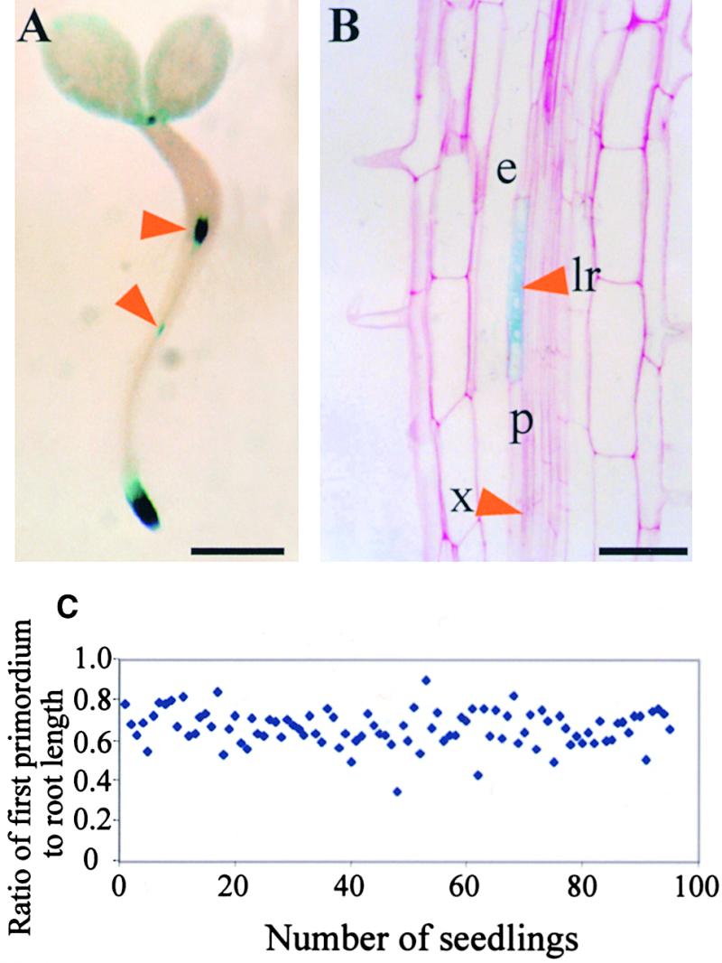
Lateral root development in Arabidopsis provides a model for the study of hormonal signals that regulate postembryonic organogenesis in higher plants. Lateral roots originate from pairs of pericycle cells, in several cell files positioned opposite the xylem pole, that initiate a series of asymmetric, transverse divisions. The auxin transport inhibitor N-1-naphthylphthalamic acid (NPA) arrests lateral root development by blocking the first transverse division(s). We investigated the basis of NPA action by using a cell-specific reporter to demonstrate that xylem pole pericycle cells retain their identity in the presence of the auxin transport inhibitor. However, NPA causes indoleacetic acid (IAA) to accumulate in the root apex while reducing levels in basal tissues critical for lateral root initiation. This pattern of IAA redistribution is consistent with NPA blocking basipetal IAA movement from the root tip. Characterization of lateral root development in the shoot meristemless1 mutant demonstrates that root basipetal and leaf acropetal auxin transport activities are required during the initiation and emergence phases, respectively, of lateral root development.


Figures

Figure 1.
Figure 1.
Developmental Stages during Lateral Root Formation in Arabidopsis. (A) Radial organization of the Arabidopsis primary root showing the cortical (C), endodermal (E), epidermal (EP), and pericycle (P) cell layers. (B) Radial section of the primary root showing a lateral root primordium (LR) developing opposite the xylem pole. (C) Longitudinal section of the primary root, with arrows indicating the cell walls of a founder pericycle cell before the first anticlinal division, initiating the formation of a lateral root primordium (stage 0). (D) Two founder pericycle cells, each having undergone a single asymmetric anticlinal division (stage Ib primordium). Arrowheads indicate the positions of the newly formed cell walls. (E) One founder cell has undergone three asymmetric anticlinal divisions, whereas the other has formed a single anticlinal division (stage Id). (F) A stage II primordium that had previously undergone three asymmetric anticlinal divisions, with the newly formed inner layer (IL) and outer layer (OL) indicated. (G) A stage II primordium that underwent six asymmetric anticlinal divisions before the first periclinal division. (H) A newly emerged lateral root primordium. formula image. Asterisks indicate xylem poles.
Figure 2.
Figure 2.
Xylem Pole Pericycle Cell Identity Is Not Altered by NPA. The GAL4-GFP enhancer trap line J0121 was grown on Murashige and Skoog agar for 7 days, and optical sections of GFP expression were imaged using multiphoton microscopy. (A) GFP was expressed within three adjacent pericycle cell files in J0121 roots. (B) A median longitudinal optical section showing that GFP expression was detected adjacent to both xylem poles. (C) A median longitudinal optical section showing that seedlings grown in the presence of 10 μM NPA also show GFP expression within files of pericycle cells adjacent to the xylem pole. formula image.
Figure 3.
Figure 3.
NAA Can Rescue the Block in Lateral Root Formation Caused by NPA. Seedlings were grown on Murashige and Skoog agar containing either 0 μM NPA (columns A and B) or 5 μM NPA (columns C and D) for 9 days before being transferred to fresh Murashige and Skoog agar with no additions (column A) or containing 0.1 μM NAA (column B), 5 μM NPA (column C), or 5 μM NPA and 0.1 μM NAA (column D). The seedlings were allowed to grow for an additional 3 days, and the total number of roots was counted. Error bars represent the sd (formula image).
Figure 4.
Figure 4.
NPA Causes a Redistribution of Root Tip IAA. The amount of IAA was measured for three segments of the primary root between 0 and 3 mm, 3 and 10 mm, and 10 and 20 mm for seedlings grown in the presence of 0, 1, 5, or 10 μM NPA. Results are shown as pg IAA/μg root tissue. Error bars represent the sd (n = 3 to 4 pooled samples of 50 to 100 root segments). Student's t test was performed on the data to demonstrate the significance of the differences observed between IAA levels in the root tip/meristem/elongation zones and the next analyzed section (formula image) and after application of 10 μM NPA (formula image).
Figure 5.
Figure 5.
The DR5::uidA Marker Detects IAA Accumulation Close to the Tip of NPA-Treated Arabidopsis Roots. (A) Expression of the synthetic auxin-responsive reporter DR5::uidA is localized to the primary root apex of 2-day-old seedlings grown in the absence of NPA. (B) to (E) Differential interference contrast images of GUS-stained DR5::uidA root apical tissues grown in the absence of NPA (B) or in the presence of 1 μM NPA (C), 5 μM NPA (D), or 10 μM NPA (E). formula image.
Figure 6.
Figure 6.
The cycB1:1::uidA Marker Reveals a Close Relationship between the Position of the First Division of Lateral Root Formation and the Root Tip. (A) Forty-hour-old seedlings containing the cycB1:1::uidA transgene were histochemically stained for GUS activity. The top arrowhead indicates staining at the transition zone between the hypocotyl and the root, and the bottom arrowhead indicates a stained lateral root primordium. formula image. (B) A 5-μm section through a stage II primordium of a 40-hour-old seedling carrying the cycB1:1::uidA transgene that was histochemically stained for GUS activity. Arrowheads indicate the lateral root primordium (lr) and the xylem vessel (x); also indicated are the pericycle cell layer (p) and the endodermis (e). formula image. (C) Seedlings (cycB1:1::uidA) were harvested every day for 7 days and histochemically stained for GUS activity. Primary root length and the distance to the root tip from the first lateral root primordium were measured in roots containing a single primordium. The ratio of the distance between the root tip and the first primordium to the length of the root is plotted (formula image).
Figure 7.
Figure 7.
stm1 Forms a Similar Number of Lateral Roots to Wild Type in Response to Auxin but Has an Altered Acropetal Development Profile. (A) Wild-type (Landsberg erecta) and stm1 plants were grown for 10 days on Murashige and Skoog agar containing 0, 0.01, or 0.1 μM NAA. The number of lateral roots per millimeter of primary root was counted for 15 seedlings under each condition. Error bars represent the sd. (B) and (C) Wild-type (B) and stm1 (C) seedlings were grown on Murashige and Skoog agar for 10 days, and the length of each lateral root and its distance from the hypocotyl–root junction were measured (formula image). The values obtained were illustrated graphically using Excel software, and the trend lines (solid bars) were calculated, indicating the presence of an acropetal lateral root developmental gradient in the wild type (B) that is absent in stm1 (C).

References

    1. Beeckman, T., and Engler, G. (1994). An easy technique for the clearing of histochemically stained plant tissue. Plant Mol. Biol. Rep. 12, 37–42.
    1. Beemster, G.T.S., and Baskin, I.S. (1998). Analysis of cell division and elongation underlying the developmental acceleration of root growth in Arabidopsis thaliana. Plant Physiol. 116, 1515–1526. - PMC - PubMed
    1. Bennett, M.J., Marchant, A., May, S.T., and Swarup, R. (1998). Going the distance with auxin: Unravelling the molecular basis of auxin transport. Philos. Trans. R. Soc. Lond. B 353, 1511–1515. - PMC - PubMed
    1. Blakely, L.M., Durham, M., Evans, T.A., and Blakely, R.M. (1982). Experimental studies on lateral root formation in radish seedling roots. I. General methods, developmental stages, and spontaneous formation of laterals. Bot. Gaz. 143, 341–352.
    1. Casero, P.J., Casimiro, I., Rodríguez-Gallardo, L., Martín-Partido, G., and Lloret, P.G. (1993). Lateral root initiation by means of asymmetrical transversal divisions of the pericycle cells in adventitious roots of Allium cepa. Protoplasma 176, 138–144.

Publication types

MeSH terms

LinkOut - more resources