. 2001 Apr;13(4):853-71.
doi: 10.1105/tpc.13.4.853.

The TRANSPARENT TESTA12 gene of Arabidopsis encodes a multidrug secondary transporter-like protein required for flavonoid sequestration in vacuoles of the seed coat endothelium

Affiliations

The TRANSPARENT TESTA12 gene of Arabidopsis encodes a multidrug secondary transporter-like protein required for flavonoid sequestration in vacuoles of the seed coat endothelium

I Debeaujon et al. Plant Cell. 2001 Apr.

Abstract

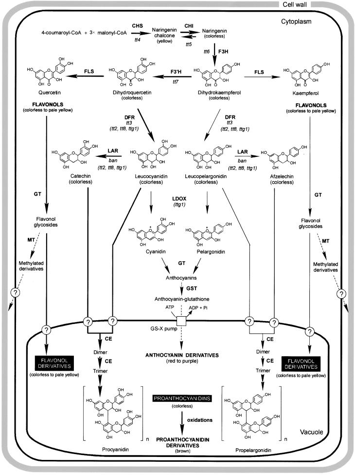
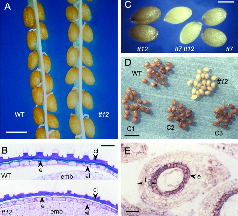
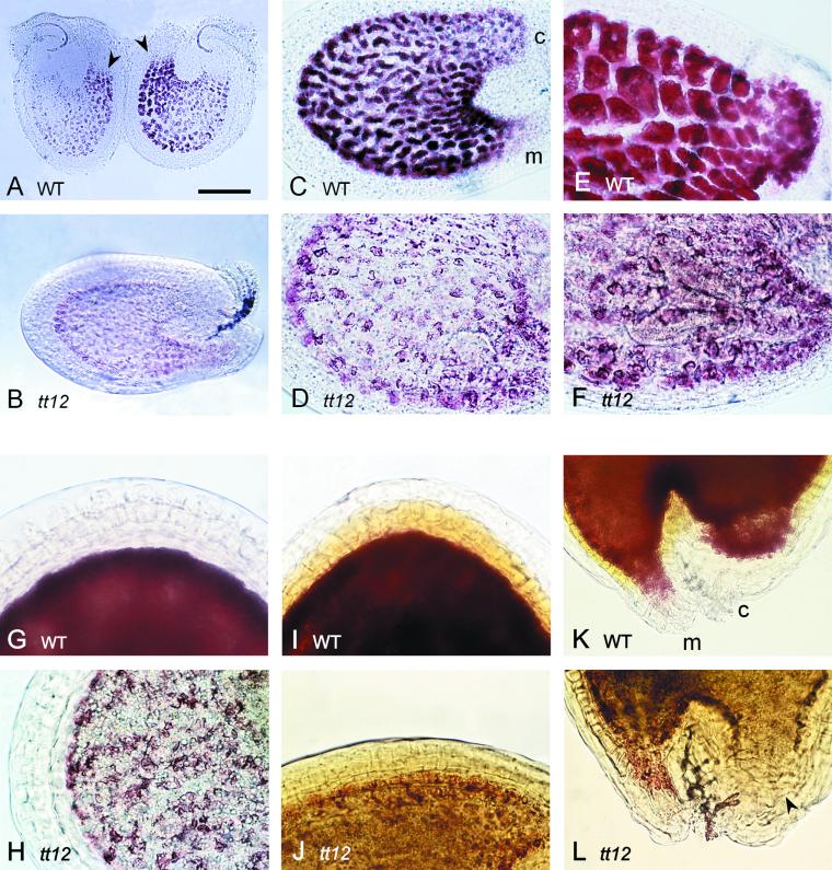
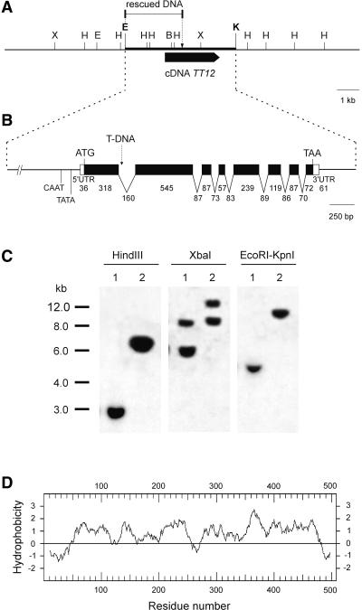
Phenolic compounds that are present in the testa interfere with the physiology of seed dormancy and germination. We isolated a recessive Arabidopsis mutant with pale brown seeds, transparent testa12 (tt12), from a reduced seed dormancy screen. Microscopic analysis of tt12 developing and mature testas revealed a strong reduction of proanthocyanidin deposition in vacuoles of endothelial cells. Double mutants with tt12 and other testa pigmentation mutants were constructed, and their phenotypes confirmed that tt12 was affected at the level of the flavonoid biosynthetic pathway. The TT12 gene was cloned and found to encode a protein with similarity to prokaryotic and eukaryotic secondary transporters with 12 transmembrane segments, belonging to the MATE (multidrug and toxic compound extrusion) family. TT12 is expressed specifically in ovules and developing seeds. In situ hybridization localized its transcript in the endothelium layer, as expected from the effect of the tt12 mutation on testa flavonoid pigmentation. The phenotype of the mutant and the nature of the gene suggest that TT12 may control the vacuolar sequestration of flavonoids in the seed coat endothelium.


Figures

Figure 1.
Figure 1.
Scheme of the Flavonoid Biosynthetic Pathway. The subpathway leading to the formation of flavonoid pigments in the Arabidopsis seed coat is indicated by boldface arrows; thin arrows represent an alternative subpathway that functions predominantly in Arabidopsis vegetative parts. The pathways to polymethylated flavonols (Chrysosplenium) and to anthocyanin vacuolar transport (maize) have not been found to occur in Arabidopsis and are thus hypothetical (dashed arrows). Enzymatic steps affected in tt mutants are indicated, with mutants corresponding to regulatory genes given in parentheses and the other mutants corresponding to structural genes. Enzymes are shown in boldface letters. CE, condensing enzyme; CHI, chalcone isomerase; CHS, chalcone synthase; DFR, dihydroflavonol-4-reductase; F3H, flavonol 3-hydroxylase; F3′H, flavonol 3′-hydroxylase; FLS, flavonol synthase; GST, glutathione S-transferase; GS-X, glutathione conjugate; GT, glycosyltransferase; LAR, leucoanthocyanidin reductase; LDOX, leucoanthocyanidin dioxygenase; MT, methyltransferase.
Figure 2.
Figure 2.
Seed Phenotypes. Mature seed subtle color nuances (as they appear in [A], [C], and [D]) are likely to vary slightly with the conditions of plant culture, the age of seed lots, and photograph conditions. These conditions were kept identical for all genotypes within each photo but differ among photos, which limits the comparability of panels (A), (C), and (D). (A) Mature seeds of the wild-type (WT) and the tt12 mutant. The tt12 mutant seeds, which have lost the bright brown-orange color characterizing wild-type seeds, are dull pale brown. Bar = 250 μm. (B) Cytochemical localization of polyphenolic compounds (in blue-green; arrowheads) in the testa of mature wild-type and tt12 seeds after staining with toluidine blue O. al, aleurone layer; cl, four crushed parenchymatic layers of the testa; e, endothelium layer of the testa; emb, embryo. Bar = 40 μm. (C) Mature seeds of the tt7 and tt12 single mutants, and of the tt7 tt12 double mutant. Bar = 250 μm. (D) Molecular complementation of the tt12 mutation. Freshly harvested seed lots after ripening for 6 months in anoxia (under Scotch tape on a slide) are presented. The seeds of three independent complementing transformants (C1 to C3) show a wild-type brown color, unlike tt12 seeds, which stayed very pale brown. Bar = 1 mm. (E) Expression of the TT12 gene in the endothelium layer of a wild-type immature seed at the globular stage of embryo development. The section was hybridized with a digoxigenin-labeled antisense TT12 probe and observed with a light microscope. The signal is dark pink (arrowhead). e, endothelium layer of the testa; t, testa. Bar = 40 μm.
Figure 3.
Figure 3.
Genetic Determinism of the Reduced Dormancy Phenotype in the tt12 Mutant. The time course of germination for freshly harvested seeds (7 days of dry storage) is presented for the wild type (WT; Ws-1), tt12, and F1 progeny from reciprocal crosses. The parent mentioned first is the female parent.
Figure 4.
Figure 4.
Localization of the tt12 Mutation on the Genetic Map of Chromosome 3. The genomic region corresponds to the bottom of chromosome 3, with “Top” indicating chromosome orientation. The genetic distance in centimorgans (cM) between two mapped markers is indicated below the given interval, plus or minus the standard error. The map is anchored by the gl1 marker position (Koornneef, 1994). The other marker positions are deduced from our own linkage data combined with the genetic map described by Koornneef (1994). The position of tt6 to the left of tt5 was deduced from the molecular mapping data of Camilleri et al. (1998).
Figure 5.
Figure 5.
Proanthocyanidin Pigment Deposition in Developing Seeds of the Wild Type and the tt12 Mutant Analyzed with Vanillin Staining. Immature seeds were incubated on a slide in vanillin HCl before observation with a microscope. Ovules and seeds younger than the two-cell stage of embryo development are not shown because no pigment was detected. The wild-type seeds (upper photo lines) are compared with tt12 mutant seeds at the same developing stage (lower photo lines). (A) and (B) Developing wild-type seeds at approximately the four-cell stage of embryo development (ED). PAs and precursors appear dark red. Arrowheads indicate the micropylar areas where pigment was first localized before spreading over the entire endothelium layer. (B) Corresponding stage in tt12. (C) and (D) Wild-type seed at the late globular stage of ED. The endothelium layer is completely pigmented, which allows distinction of the micropyle from the chalaza, but vacuoles are not completely filled up. (D) Corresponding stage in tt12. (E) and (F) Wild-type seed at the heart stage of ED. The chalazal bulb shows large endothelial cells filled with pigments. (F) Corresponding stage in tt12. (G) and (H) Wild-type seed at the torpedo stage of ED. (H) Corresponding stage in tt12. (I) through (L) Wild-type seed at the walking stick–cotyledonary stage of ED. A second pigment is spreading from the endothelium layer to the upper parenchymal cell layers. (I) and (K) show the seed region opposite the chalaza–micropyle pole and the chalaza–micropyle areas, respectively. (J) and (L) Corresponding stages in tt12. c, chalaza; m, micropyle; WT, wild type. Bar in (A) = 75 μm for (A), 60 μm for (B), 50 μm for (C), and 30 μm for (D) to (L).
Figure 6.
Figure 6.
Organization of the Genomic Region and Structure of the TT12 Gene. (A) Localization of TT12 cDNA on the restriction map of a λ clone. The arrow indicates the sense of transcription. The genomic DNA fragment isolated by plasmid rescue is shown. The genomic fragment used to complement the tt12 mutation is indicated by a thick line. Restriction sites are as follows: B, BamHI; E, EcoRI; H, HindIII; K, KpnI; and X, XbaI. (B) Sequence features of the TT12 gene. Boxes represent exons, with white boxes indicating the cDNA untranslated regions (UTRs). The arrow indicates the site of T-DNA insertion in the tt12 mutant. Sizes are drawn to scale. The entire TT12 cDNA sequence was submitted to GenBank under the accession number AJ294464. (C) DNA gel blot analysis of wild-type and tt12 plants. Restricted genomic DNA of wild-type (lanes 1) and tt12 (lanes 2) plants probed with the complete cDNA reveals a polymorphism between wild type and tt12. The positions and lengths (in kilobases) of DNA molecular mass markers are indicated at left. (D) Hydropathy profile of the TT12 protein, as determined by the method of Kyte and Doolittle (1982), using a window of 19 amino acid residues.
Figure 7.
Figure 7.
Alignment of TT12 with Five Related Proteins. The alignment involves two close paralogs of TT12 and the three orthologous proteins from the MATE family that were previoulsy characterized. The six sequences were aligned using the CLUSTALW program with default parameters. Identical and similar residues are shown on backgrounds of black and gray, respectively. Gaps required for optimal alignment are indicated by dashes. The putative TMs of the TT12 protein, as determined by the TMHMM program, are delimited by thin lines above the sequences; outer (O) and inner (I) hydrophilic internal segments also are indicated. A comparison of TT12 with its 30 Arabidopsis paralogs was performed (data not shown); residues conserved in all sequences are indicated by stars, and those conserved in all but one sequence are indicated by dots. Triangles indicate the charged residues present in TMs. Conserved protein domains (D1 to D5) are represented by thick lines. Ath, Arabidopsis; Sce, S. cerevisiae; Vpa, V. parahaemolyticus; Eco, E. coli.
Figure 8.
Figure 8.
Detection of the TT12 mRNA by Quantitative RT-PCR. Results of 21-cycle PCR amplifications are presented. The Arabidopsis polyubiquitin gene UBQ10 was used as a loading control. (A) Detection in diverse tissues from wild-type plants. RNA preparations were made from 4-day-old seedlings (Sg), rosette leaves (L), stems (St), roots (R), buds (B), flowers (F), and immature siliques (stages 1 to 9; see Methods for descriptions). (B) Detection in immature siliques (stage 3) of wild-type (lane 1) and tt12 (lane 2) plants.

Similar articles

Cited by

References

    1. Albert, S., Delseny, M., and Devic, M. (1997). BANYULS, a novel negative regulator of flavonoid biosynthesis in the Arabidopsis seed coat. Plant J. 11, 289–299. - PubMed
    1. Alfenito, M.R., Souer, E., Goodman, C.D., Buell, R., Mol, J., Koes, R., and Walbot, V. (1998). Functional complementation of anthocyanin sequestration in the vacuole by widely divergent glutathione S-transferases. Plant Cell 10, 1135–1149. - PMC - PubMed
    1. Barz, W. (1977). Degradation of polyphenols in plants and plant cell suspension cultures. Physiol. Veg. 15, 261–277.
    1. Baur, P.S., and Walkinshaw, C.H. (1974). Fine structure of tannin accumulation in callus cultures of Pinus elliotti (slash pine). Can. J. Bot. 52, 615–619.
    1. Bechtold, N., Ellis, J., and Pelletier, G. (1993). In planta Agrobacterium-mediated gene transfer by infiltration of adult Arabidopsis thaliana plants. C. R. Acad. Sci. Paris 316, 1194–1199.

Publication types

MeSH terms