

Spatial and temporal independent component analysis of functional MRI data containing a pair of task-related waveforms
- PMID: 11284046
- PMCID: PMC6871956
- DOI: 10.1002/hbm.1024
Spatial and temporal independent component analysis of functional MRI data containing a pair of task-related waveforms
Erratum in
-
Correction to "Spatial and temporal independent component analysis of functional MRI data containing a pair of task-related waveforms".Hum Brain Mapp. 2024 Aug 15;45(12):e70007. doi: 10.1002/hbm.70007. Hum Brain Mapp. 2024. PMID: 39189682 Free PMC article. No abstract available.
Abstract
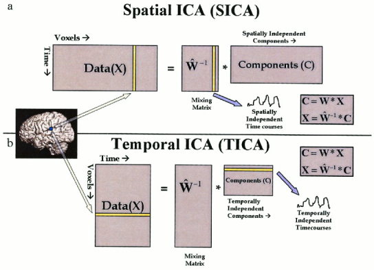
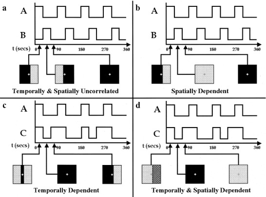
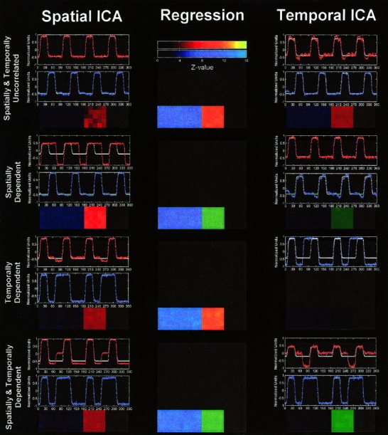
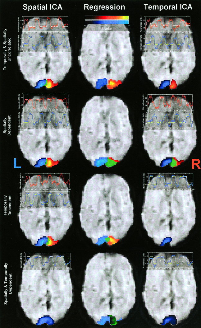
Independent component analysis (ICA) is a technique that attempts to separate data into maximally independent groups. Achieving maximal independence in space or time yields two varieties of ICA meaningful for functional MRI (fMRI) applications: spatial ICA (SICA) and temporal ICA (TICA). SICA has so far dominated the application of ICA to fMRI. The objective of these experiments was to study ICA with two predictable components present and evaluate the importance of the underlying independence assumption in the application of ICA. Four novel visual activation paradigms were designed, each consisting of two spatiotemporal components that were either spatially dependent, temporally dependent, both spatially and temporally dependent, or spatially and temporally uncorrelated, respectively. Simulated data were generated and fMRI data from six subjects were acquired using these paradigms. Data from each paradigm were analyzed with regression analysis in order to determine if the signal was occurring as expected. Spatial and temporal ICA were then applied to these data, with the general result that ICA found components only where expected, e.g., S(T)ICA "failed" (i.e., yielded independent components unrelated to the "self-evident" components) for paradigms that were spatially (temporally) dependent, and "worked" otherwise. Regression analysis proved a useful "check" for these data, however strong hypotheses will not always be available, and a strength of ICA is that it can characterize data without making specific modeling assumptions. We report a careful examination of some of the assumptions behind ICA methodologies, provide examples of when applying ICA would provide difficult-to-interpret results, and offer suggestions for applying ICA to fMRI data especially when more than one task-related component is present in the data.
Copyright 2001 Wiley-Liss, Inc.
Figures
References
-
- Bell AJ, Sejnowski TJ (1995): An information maximisation approach to blind separation and blind deconvolution. Neural Computation 7: 1129–1159. - PubMed
-
- Biswal BB, Ulmer JL (1999): Blind source separation of multiple signal sources of fMRI data sets using independent component analysis. J Comput Assist Tomogr 23: 265–271. - PubMed
-
- Biswal BB, Van Kylen J, Hyde JS (1997): Simultaneous assessment of flow and BOLD signals in resting‐state functional connectivity maps. NMR Biomed 10: 165–170. - PubMed
-
- Calhoun V, Golay X, Pearlson G (2000a): Improved fMRI slice timing correction: interpolation errors and wrap around effects. In: Proceedings ISMRM, Ninth Annual Meeting, Denver.
MeSH terms
LinkOut - more resources
Full Text Sources
Other Literature Sources
Miscellaneous
