. 2001 Apr 2;20(7):1681-91.
doi: 10.1093/emboj/20.7.1681.

Molecular basis of thermosensing: a two-component signal transduction thermometer in Bacillus subtilis

Affiliations

Molecular basis of thermosensing: a two-component signal transduction thermometer in Bacillus subtilis

P S Aguilar et al. EMBO J. .

Abstract

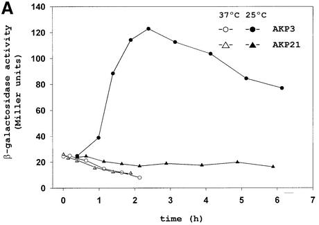
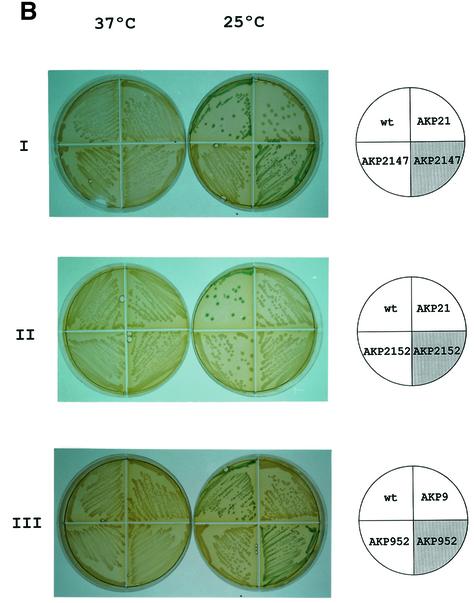
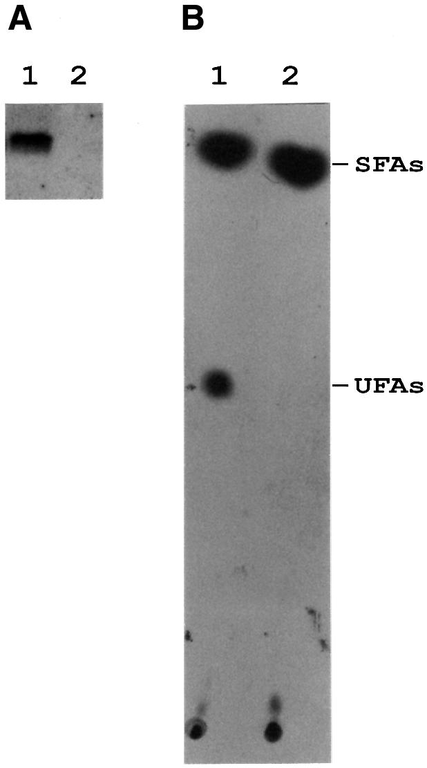
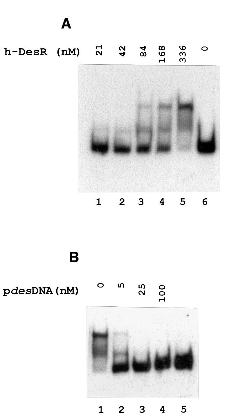
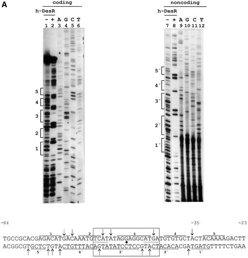
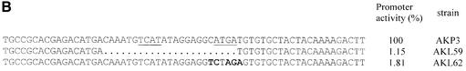
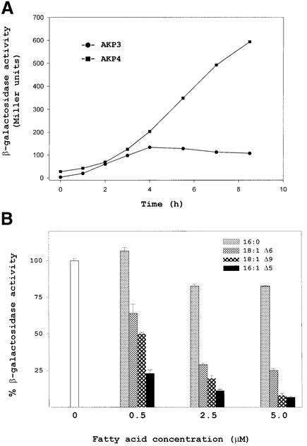
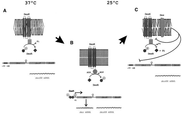
Both prokaryotes and eukaryotes respond to a decrease in temperature with the expression of a specific subset of proteins. Although a large body of information concerning cold shock-induced genes has been gathered, studies on temperature regulation have not clearly identified the key regulatory factor(s) responsible for thermosensing and signal transduction at low temperatures. Here we identified a two-component signal transduction system composed of a sensor kinase, DesK, and a response regulator, DesR, responsible for cold induction of the des gene coding for the Delta5-lipid desaturase from Bacillus subtilis. We found that DesR binds to a DNA sequence extending from position -28 to -77 relative to the start site of the temperature-regulated des gene. We show further that unsaturated fatty acids (UFAs), the products of the Delta5-desaturase, act as negative signalling molecules of des transcription. Thus, a regulatory loop composed of the DesK-DesR two-component signal transduction system and UFAs provides a novel mechanism for the control of gene expression at low temperatures.


Figures

None
Fig. 1. Pattern of des–lacZ expression in wild-type and yocFyocG cells before and after temperature downshift. (ABacillus subtilis AKP3 cells (circles) and AKP21 cells (triangles) harbouring a des–lacZ transcriptional fusion were grown at 37°C to an optical density of 0.35 (at 525 nm) and then divided into two samples. One sample was transferred to 25°C (filled symbols), and the other was kept at 37°C (open symbols). Specific β-gal activities were determined at the indicated time intervals. (B) Bacterial strains [wild-type AKP3 (wt), yocFyocG (AKP21), yocFyocG pXyl:yocFG (AKP2147), yocFyocG pXyl:yocG (AKP2152), yocG (AKP9) and yocG pXyl:yocG (AKP952)] were streaked onto Luria–Bertani (LB) medium containing 30 µg/µl X-Gal, with (shaded quarter) or without (empty quarter) the addition of 0.8% l-xylose. The strains were incubated at 37°C for 12 h (left column) or for 5 h at 37°C, and then transferred to 25°C for 36 h (right column) before photography.
None
Fig. 2. des mRNA and UFA production in wild-type and yocG strains after a downshift temperature. (A) Northern blot analysis using formaldehyde agarose gels was carried out as described in Materials and methods. Total RNA was isolated from strains JH642 (lane 1) or strain AKP8 (yocG, lane 2) grown until mid-exponential phase at 37°C and then shifted to 25°C by 30 min. Each lane contains 10 µg of total RNA. (B) Fatty acids synthesized by strains JH642 and AKP8 at 25°C. Cultures of strains JH642 (lane 1) and AKP8 (lane 2) were grown to mid-exponential phase at 37°C, then 2 ml of these cultures were challenged with 10 µCi of [14C]acetate and further shifted to 25°C for 12 h. The lipids were then extracted and transesterified, and the resulting methyl esters were separated into saturated (SFAs) and unsaturated (UFAs) fractions by chromatography on 20% silver nitrate-impregnated silica gel thin-layers plates. The plates were developed at –17°C and autoradiographed by 7 days. The sample in lane 1 contained 15 000 c.p.m. and 2000 c.p.m. in the SFA and UFA fractions, respectively. The sample in lane 2 contained 14 000 c.p.m. in the SFA fraction, while the UFA fraction contained only background levels of radioactivity.
None
Fig. 3. Gel shift assay showing the binding of DesR to the des promoter region. (A) The 367 bp des promoter fragment (pdesDNA) was prepared by [α-32P]dATP PCR labelling as described in Materials and methods. The [32P]pdesDNA concentration in the binding mixtures was 1.7 nM in all cases. The concentration of h-DesR used in each binding reaction is indicated above the respective line. (B) Specific competition in binding reactions using 1.7 nM [32P]pdesDNA and 336 nM h-DesR. Lane 1 shows the retarded species in the absence of unlabelled homologous DNA. Lanes 2, 3 and 4 show the dissociation of the labelled complex in the presence of 3, 15 and 60-fold molar excess of unlabelled pdesDNA, respectively, added to the binding mixtures before the addition of h-DesR. Lane 5 shows [32P]pdesDNA without the addition of h-DesR.
None
Fig. 4. DNase I footprinting assay of the des promoter region and in vivo characterization of the des promoter protected region. (A) DNase I footprinting of h-DesR protein on both strands of a 178 bp DNA fragment containing the des promoter (see Materials and methods). Sequencing reactions were performed on the same DNA fragment labelled at the coding (lanes 3 to 6) and non-coding (lanes 9 to 12) strands. Lanes 1, 2, 7 and 8 show the DNase I digestion products of pdesDNA in the presence (+) or absence (–) of h-DesR. Brackets mark the protected regions in each strand. The putative 17 bp symmetric region of the protected region is boxed, with the dyad axis of symmetry indicated by a dot. The inverted repeats are underlined with dots. DNase I footprints on both strands are shown. Arrows indicate hypersensitive bonds. (B) Promoter mutations. The sequence changes in the promoter variants are depicted along the protected region of the des promoter. The deleted region is indicated by dots, and the nucleotide changes to introduce mutations in the left inverted repeat are shown in bold characters. The inverted repeat sequences are underlined. The strains were grown at 37°C to an OD of 0.30 and then subjected to a downshift to 25°C. After 3 h of growth at 25°C, the cells were harvested and β-gal activities were determined. The average value of β-gal activity of strain AKP3 (bearing the wild-type promoter) was taken as 100% of promoter activity. The results shown are the average of three independent experiments.
None
Fig. 5. Overexpression of DesR leads to constitutive expression of des. (A) JH642 cells (wild type) were cultured at 37°C until mid-exponential phase (lane 1) and then shifted to 25°C for 30 min (lane 2) or 3 h (lane 3). Samples were taken and cell extracts were analysed for the presence of DesR with DesR antiserum. AKP2152 (2152) cells were cultured at 37°C in the absence (lane 4) or presence of 0.8% l-xylose (lane 5) to mid-exponential phase and then shifted to 25°C by 2 h. Samples were taken and analysed for the presence of DesR as described above. (B) Strains JH642 (wt, lane 1), AKP2152 (2152) (lanes 2 and 3), AKP2047 (2047) (lanes 4 and 5) and AKP20 (20) (lane 6) were cultured at 37°C until mid-exponential phase and then shifted to 25°C by 2 h. AKP2152 and AKP2047 cultures were supplemented (+) or not (–) with 0.8% xylose. Samples were taken and proteins from the same quantity of cells were analysed for the presence of DesR. (C) Bacterial strains AKP3 (wt), AKP20 (desK,pkan-desR) and AKP2047 (AKP20 pXyl-desKdesR::thr) were streaked onto LB medium containing 30 µg/µl X-Gal, with (shaded quarter) or without (empty quarter) the addition of 0.8% l-xylose. The strains were incubated at 37°C for 12 h (left) or for 5 h at 37°C, and then transferred to 25°C for 36 h (right) before photography.
None
Fig. 6. desKR mRNA levels before and after a downshift temperature. Northern blot analysis using formaldehyde agarose gels was carried out as described in Materials and methods. Total RNA was isolated from strain JH642 grown until mid-exponential phase at 37°C (lane 1) and then shifted to 25°C by 30 min (lane 2) or 180 min (lane 3). Each lane contains 15 µg of total RNA.
None
Fig. 7. The expression of the des gene is regulated by UFAs. (ABacillus subtilis strains AKP3 (des+) (circles) and AKP4 (des) (squares) harbouring a des–lacZ fusion located in the amyE locus were grown at 37°C to an OD of 0.35 and then transferred to 25°C. Aliquots were taken and specific β-gal activities were determined at the indicated time intervals. (BBacillus subtilis AKP4 cells were grown at 37°C to an OD of 0.27 at 525 nm and were then subjected to 0, 0.5, 2.5 or 5 µM of the indicated fatty acids before being transferred to 25°C. After 4 h of growth at 25°C, the cells were harvested and β-gal activities were determined. The average value of β-gal activity without supplement of fatty acid was taken as 100% of activity. The results shown are the average of three independent experiments.
None
Fig. 8. Model of des transcriptional control by two-component temperature signal transduction proteins. It is proposed that DesK assumes different signalling states in response to a temperature-induced change in membrane fluidity. This is accomplished by regulating the ratio of kinase to phosphatase activity such that a phosphatase-dominant state is present at 37°C, when membrane lipids are disordered (A), whereas a kinase-dominant state predominates upon an increase in the proportion of ordered membrane lipids after a temperature downshift to 25°C (B). DesK-mediated phosphorylation of DesR results in transcriptional activation of des (B). Activation of des results in synthesis of Des, which desaturates the acyl chains of membrane phospholipids (C). These newly synthesized UFAs inhibit des transcription either by favouring DesK dephosphorylation of DesR-P or by causing dissociation of DesR-P from its binding site (C) (see text for further details).

Similar articles

Cited by

References

    1. Aguilar P.S., Cronan,J.E.,Jr and de Mendoza,D. (1998) A Bacillus subtilis gene induced by cold-shock encodes a membrane phospholipid desaturase. J. Bacteriol., 180, 2194–2200. - PMC - PubMed
    1. Aguilar P.S., Lopez,P. and de Mendoza,D. (1999) Transcriptional control of the low-temperature-inducible des gene, encoding the Δ5 desaturase of Bacillus subtilis. J. Bacteriol., 181, 7028–7033. - PMC - PubMed
    1. Choi J.Y., Stukey,J., Hwang,S.Y. and Martin,C.E. (1996) Regulatory elements that control transcription activation and unsaturated fatty acid-mediated repression of the Saccharomyces cerevisiae OLE1 gene J. Biol. Chem., 271, 3581–3589. - PubMed
    1. Cronan J.E. Jr and Rock,C.O. (1996) Biosynthesis of membrane lipids. In Neidhart,F.C. et al. (eds), Escherichia coli and Salmonella typhimurium: Cellular and Molecular Biology. 2nd edn, Vol. 1. American Society for Microbiology, Washington, DC, pp. 612–636.
    1. Dartois V., Débarbouillé,F., Kunst,F. and Rapaport,G. (1998) Characterization of a novel member of the DegS–DegU regulon affected by salt stress in Bacillus subtilis. J. Bacteriol., 180, 1855–1861. - PMC - PubMed

Publication types

MeSH terms

LinkOut - more resources