. 2001 Apr 2;20(7):1715-25.
doi: 10.1093/emboj/20.7.1715.

Transcriptional repression and developmental functions of the atypical vertebrate GATA protein TRPS1

Affiliations

Transcriptional repression and developmental functions of the atypical vertebrate GATA protein TRPS1

T H Malik et al. EMBO J. .

Abstract

Known vertebrate GATA proteins contain two zinc fingers and are required in development, whereas invertebrates express a class of essential proteins containing one GATA-type zinc finger. We isolated the gene encoding TRPS1, a vertebrate protein with a single GATA-type zinc finger. TRPS1 is highly conserved between Xenopus and mammals, and the human gene is implicated in dominantly inherited tricho-rhino-phalangeal (TRP) syndromes. TRPS1 is a nuclear protein that binds GATA sequences but fails to transactivate a GATA-dependent reporter. Instead, TRPS1 potently and specifically represses transcriptional activation mediated by other GATA factors. Repression does not occur from competition for DNA binding and depends on a C-terminal region related to repressive domains found in Ikaros proteins. During mouse development, TRPS1 expression is prominent in sites showing pathology in TRP syndromes, which are thought to result from TRPS1 haploinsufficiency. We show instead that truncating mutations identified in patients encode dominant inhibitors of wild-type TRPS1 function, suggesting an alternative mechanism for the disease. TRPS1 is the first example of a GATA protein with intrinsic transcriptional repression activity and possibly a negative regulator of GATA-dependent processes in vertebrate development.


Figures
None
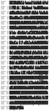
Fig. 1. TRPS1 is a highly conserved and atypical vertebrate GATA protein. Deduced amino acid sequences of Xenopus, mouse and human TRPS1. Xenopus and mouse cDNAs were isolated as described; the human sequence is based in part on confirmed EST clones and in part on recently published data (Momeni et al., 2000). Identical amino acids are shaded in black, and conservative substitutions in gray. Positions of the nine putative zinc finger motifs are indicated by arrows under the sequence, the GATA-type zinc finger (residues 886–910 in XTRPS1) is marked by a thick arrow, and the two putative nuclear localization signals are marked by shaded boxes.
None
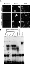
Fig. 2. TRPS1 is a sequence-specific, DNA-binding nuclear protein. (A) Subcellular localization by immunofluorescence of COS cells transfected with cDNA constructs encoding full-length XTRPS1 (top), an N-terminal truncated protein (XTRPS1ΔN, middle) or full-length mTRPS1 (bottom). Cells transfected with Xenopus or mouse proteins were stained with rabbit antisera directed against Xenopus and mouse peptides, respectively. Results with the appropriate pre-immune serum and with 4′-6-diamidine-2-phenylindole (DAPI) nuclear stain are also shown. (B) Electrophoretic mobility shift assay (EMSA) of in vitro translated XTRPS1ΔN using a double-stranded GATA oligonucleotide probe. Cold competitions are with either the same oligonucleotide or one in which the GATA sequence was altered to GATC. The complex formed with XTRPS1ΔN (arrow) is abrogated in the presence of appropriate antiserum. Full-length XGATA4, and a mutant protein in which two cysteine residues within the XTRPS1 GATA-type zinc finger are altered (XTRPS1ΔNmut), serve as additional controls.
None
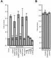
Fig. 3. TRPS1 functions as a sequence-specific transcriptional repressor. Results of transient transfection of COS cells with a GATA-dependent luciferase reporter (A) and expression constructs encoding the transcriptional activator XGATA4 either alone or in combination with constructs expressing full-length XTRPS1, the truncated protein XTRPS1ΔN, full-length mTRPS1 and the corresponding mutant proteins XTRPS1mut, XTRPS1ΔNmut and mTRPS1mut, in which the GATA-type zinc finger is disrupted. Mut reporter, a reporter carrying mutated GATA sites. Dose dependence of XTRPS1-mediated transcriptional repression was established by co-transfection of XGATA4 and XTRPS1 plasmids in the indicated ratios. (B) TRPS1 fails to repress a GATA-independent reporter that is activated by the combination of a TCF-family protein and β-catenin.
None
Fig. 4. TRPS1 represses GATA-mediated induction of endoderm in Xenopus embryos. RT–PCR analysis of the endodermal marker genes IFABP, LFABP and XGATA5 on explanted Xenopus animal caps co-injected with 400 pg of XGATA4 (A and B) or Mixer (B) mRNA and 300–600 pg of XTRPS1ΔN or 600 pg of mTRPS1 mRNA. Controls include embryos injected with H2O or with a neutral filler (Ctl) RNA, and RT–PCR analysis for EF-1α and on samples not treated with reverse transcriptase (–RT). Results are representative of four independent experiments. (C) PCR on representative samples [lanes 3 and 4 from (A)] confirms that reactions were performed in the linear range of amplification.
None
Fig. 5. The repressor domain of TRPS1 maps to a C-terminal region encompassing two Ikaros-type zinc fingers. (A) Schematic of expression constructs 1–5 used in these experiments. 1–3 are truncation mutants of XTRPS1; 4 and 5 are fusion constructs between portions of the transcriptional activator XGATA4 and the C-terminal 119 residues of XTRPS1. G-Zn and C-Zn designate the TRPS1 GATA-type zinc finger and the C-terminal zinc finger of XGATA4, respectively. (B) EMSA analysis of the proteins encoded by constructs 1–5 using a GATA probe and competitor oligonucleotides as shown in Figure 2. (C) Results of transient transfection of COS cells with a GATA-dependent luciferase reporter and either constructs 4 or 5 alone, or constructs encoding XGATA4 either alone or in combination with constructs 1–5. Mut rep, a reporter carrying mutated GATA sites. (D) RT–PCR analysis of IFABP expression in explanted Xenopus animal caps injected with H2O or co-injected with XGATA4 (400 pg) and the indicated TRPS1 (600 pg) mRNAs. (E) Comparison of the amino acid sequence of mTRPS1 residues 1204–1270 with the C-terminus of each murine Ikaros-family protein. Identical amino acid residues are shaded in black and conservative substitutions in gray.
None
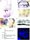
Fig. 6. Correlations between TRPS1 in development and disease. (A–E) mRNA in situ hybridization analysis of mTRPS1 expression in whole mouse embryos (A), the jaw (B, coronal section), digits (C), femoral head (D) and scalp hair follicles (E) at E12.5–13.5. Hybridization with a sense probe yielded no detectable signal (data not shown). (F) Northern analysis of mTRPS1 (T) or GAPDH (G) expression in tissues isolated from mouse fetuses at the indicated gestational age. FL, forelimbs; HL, hind limbs. (G) Map from the Jackson Laboratory BSS backcross showing a portion of chromosome 15 with loci linked to Trps1, depicted with the centromere at the top. Percentage recombination between adjacent loci (±SE) are indicated to the left and gene symbols to the right. Loci mapping to the same position are listed on the same line. Missing typings were inferred from surrounding data where assignment was unambiguous. Panel data and references for mapping other depicted loci are available at http://www.jax.org/resources/documents/cmdata. (H) FISH analysis of hTRPS1 showing localization to chromosome 8q23–24.
None
Fig. 7. TRPS1 truncation mutations associated with type I tricho-rhino-phalangeal syndrome function as dominant inhibitors of the wild-type protein. (A) Results of transient transfection of COS cells with a GATA-dependent luciferase reporter and XGATA4 plasmid, either alone (lane 2) or in combination with mTRPS1 and the disease isoforms P1–P4 (lanes 3–7). P1–P4 are mutants of mTRPS1 designed to mimic the following truncation mutations identified in TRPS patients (Momeni et al., 2000), respectively: C338X, R611X, R840X and a frameshift from codon 1121 that results in premature termination. Lanes 8–11 show that P1–P4 lack intrinsic transactivation capacity. (B) RT–PCR analysis of Xenopus IFABP, LFABP and GATA5 expression, or the controls EF-1α and mTRPS1 on Xenopus animal caps injected with H2O or co-injected with 300 pg of XGATA4 (lanes 1–6), 500 pg of mTRPS1 (lanes 4–6) and 500 pg of P1 or P3 (lanes 2–5) mRNAs.

References

    1. Cubadda Y., Heitzler,P., Ray,R.P., Bourouis,M., Ramain,P., Gelbart,W., Simpson,P. and Haenlin,M. (1997) u-shaped encodes a zinc finger protein that regulates the proneural genes achaete and scute during the formation of bristles in Drosophila. Genes Dev., 11, 3083–3095. - PMC - PubMed
    1. da Costa L.T., He,T.C., Yu,J., Sparks,A.B., Morin,P.J., Polyak,K., Laken,S., Vogelstein,B. and Kinzler,K.W. (1999) CDX2 is mutated in a colorectal cancer with normal APC/β-catenin signaling. Oncogene, 18, 5010–5014. - PubMed
    1. Deconinck A.E., Mead,P.E., Tevosian,S.G., Crispino,J.D., Katz,S.G., Zon,L.I. and Orkin,S.H. (2000) FOG acts as a repressor of red blood cell development in Xenopus. Development, 127, 2031–2040. - PubMed
    1. Fukushige T., Hawkins,M.G. and McGhee,J.D. (1998) The GATA-factor elt-2 is essential for formation of the Caenorhabditis elegans intestine. Dev. Biol., 198, 286–302. - PubMed
    1. Georgopoulos K., Winandy,S. and Avitahl,N. (1997) The role of the Ikaros gene in lymphocyte development and homeostasis. Annu. Rev. Immunol., 15, 155–176. - PubMed

Publication types

MeSH terms

Associated data