. 2001 Apr 2;20(7):1785-96.
doi: 10.1093/emboj/20.7.1785.

SC35 autoregulates its expression by promoting splicing events that destabilize its mRNAs

Affiliations

SC35 autoregulates its expression by promoting splicing events that destabilize its mRNAs

A Sureau et al. EMBO J. .

Abstract

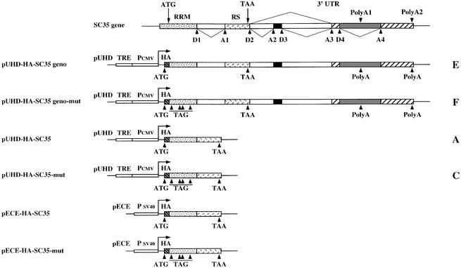
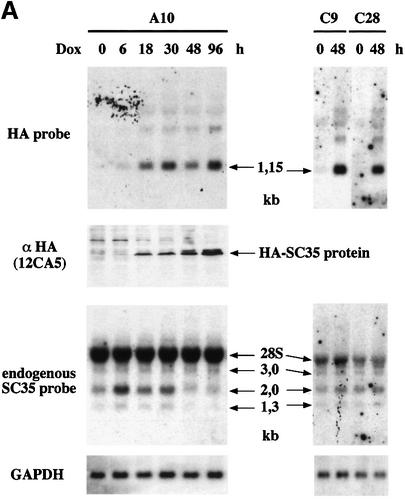
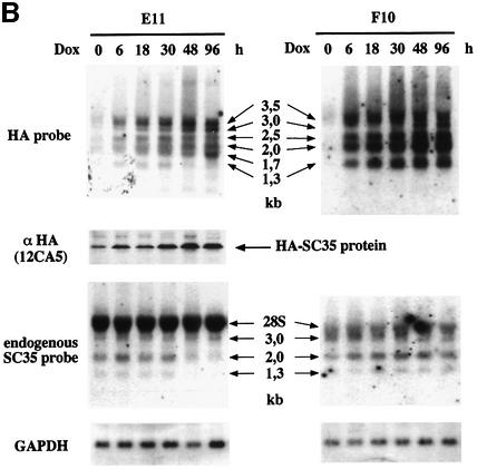
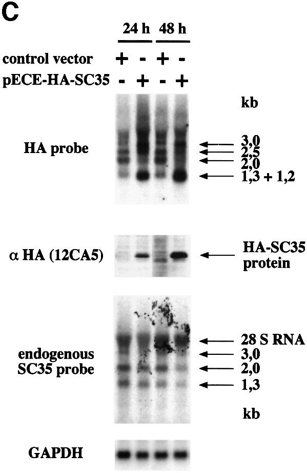
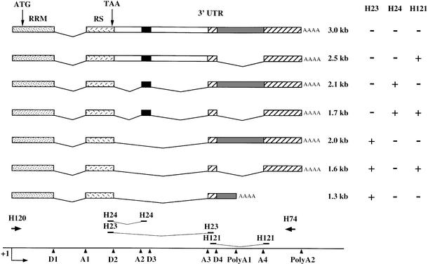
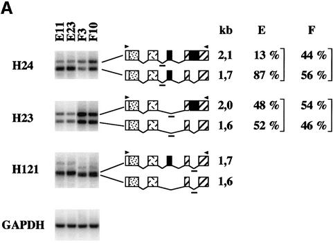
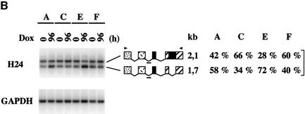
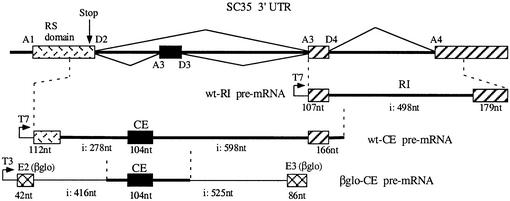
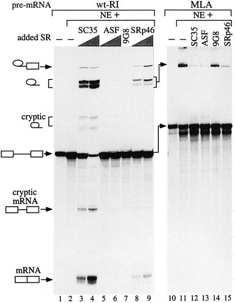
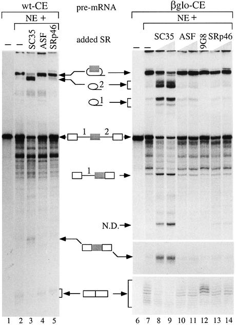
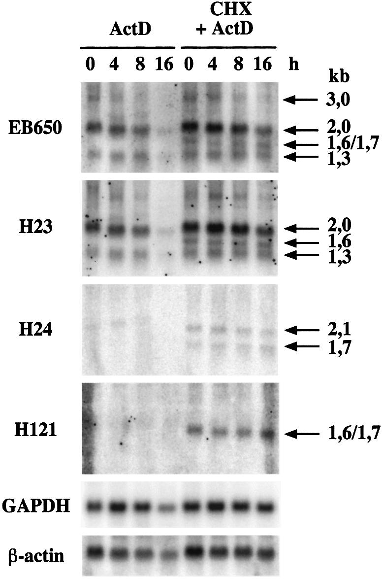
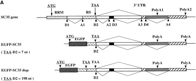
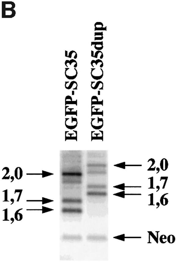
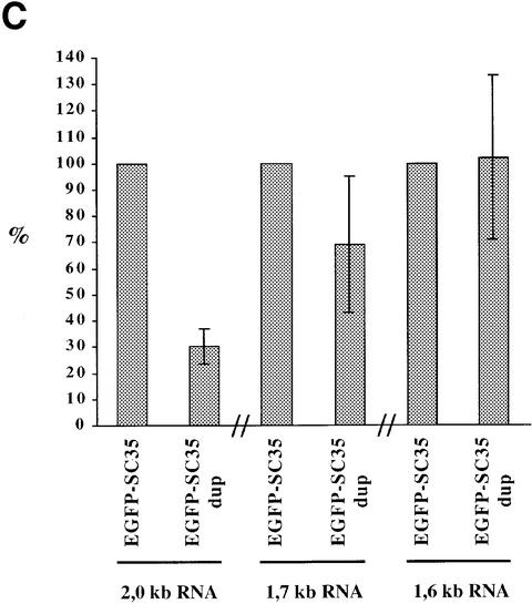
SC35 belongs to the family of SR proteins that regulate alternative splicing in a concentration-dependent manner in vitro and in vivo. We previously reported that SC35 is expressed through alternatively spliced mRNAs with differing 3' untranslated sequences and stabilities. Here, we show that overexpression of SC35 in HeLa cells results in a significant decrease of endogenous SC35 mRNA levels along with changes in the relative abundance of SC35 alternatively spliced mRNAs. Remarkably, SC35 leads to both an exon inclusion and an intron excision in the 3' untranslated region of its mRNAs. In vitro splicing experiments performed with recombinant SR proteins demonstrate that SC35, but not ASF/SF2 or 9G8, specifically activates these alternative splicing events. Interestingly, the resulting mRNA is very unstable and we present evidence that mRNA surveillance is likely to be involved in this instability. SC35 therefore constitutes the first example of a splicing factor that controls its own expression through activation of splicing events leading to expression of unstable mRNA.


Figures

None
Fig. 1. Structure of the SC35-inducible and constitutive expression vectors. Top: overall organization of the human SC35 gene. The different donor (D) and acceptor (A) splice sites as well as alternative polyadenylation sites are indicated. The two first exons encoding the SC35 RRM and RS domains are shown. Alternative splicing events occurring in the 3′UTR are represented. In the pUHD inducible vectors, the tetracyclin-transactivator responsive element (TRE), the minimal CMV promoter (PCMV) and the HA tag (HA) are indicated. In the pECE vector, SC35 expression is driven by SV40 promoter sequences (PSV40). In non-coding versions of the vectors, the in-frame ATG codons mutated into stop codons are indicated by arrowheads and TAG. The capital letter at the right of each vector corresponds to the general name of HeLa cell clones isolated following transfection with this construct.
None
Fig. 2. Northern and western blot analysis of SC35 exogenous and endogenous products in HeLa cells. (A) Analysis of SC35 expression products in HeLa cell clones stably transfected with coding (A10) or non-coding (C9 and C28) HA-tagged SC35 cDNA sequences. (B) Analysis of SC35 expression products in HeLa cell clones stably transfected with coding (E11) or non-coding (F10) HA-tagged SC35 genomic sequences. Total RNA (20 µg/lane) purified from HeLa cell clones at different times (in hours) following doxycyclin (Dox) induction was analyzed by northern blot hybridization with the HA probe specific for exogenous transcripts or with the SC35 leader probe specific for endogenous mRNAs. The size of the different SC35 mRNAs is mentioned. The 28S rRNA detected by the SC35 leader probe is indicated. Hybridization with a GAPDH-specific probe was performed to normalize for RNA amounts and transfer efficiency. Protein samples (30 µg/lane) purified from the same cells were analyzed by western blotting with the 12CA5 monoclonal antibody specific for the HA tag. (C) Analysis of SC35 expression products in F3 cells expressing non-coding HA-tagged SC35 genomic sequences and transiently transfected with either the control or pECE-HA-SC35 vector driving constitutive expression of a 1.2 kb SC35 mRNA. Northern and western blot analyses were performed as above at the indicated times following transient transfection.
None
Fig. 3. Schematic structure of the SC35 transcripts. The features of the SC35 gene are indicated as in Figure 1. For the various mRNA isoforms, whose size is indicated on the right, exons are represented by boxes and excised introns by lines. The positions of the different oligonucleotides used for PCR or Southern blot analysis of RT–PCR products (Figure 4) are indicated at the bottom. The specificity of the oligonucleotidic probes for the different SC35 mRNAs is indicated on the right.
None
Fig. 4. Southern blot analysis of RT–PCR products corresponding to exogenous and endogenous SC35 mRNAs expressed in HeLa cell clones. One-tenth of the PCR products corresponding to SC35 exogenous (A) or endogenous (B) mRNAs expressed in different HeLa cell clones were analyzed with the indicated probes. The structure and size of the transcripts corresponding to the PCR products are indicated and the positions of the PCR primers (arrowheads) and probes (short bold line) are shown. For each probe, the percentage of the hybridization signal corresponding to each PCR product was determined by PhosphorImager analysis for four independent clones of the same series and the mean values are indicated on the right. RT–PCR analysis of the GAPDH mRNA was performed to normalize for RNA amounts used in the RT step. (A) RT–PCR analysis of exogenous SC35 mRNAs from HeLa cell clones stably transfected with coding (E11 and E23) or non-coding (F3 and F10) SC35 genomic sequences and incubated for 48 h in the presence of Dox. (B) RT–PCR analysis of endogenous SC35 mRNAs expressed in HeLa cell clones stably transfected with coding cDNA (A), non-coding cDNA (C), coding genomic (E) or non-coding genomic (F) SC35 sequences prior to or 96 h after Dox induction.
None
Fig. 5. Origin and schematic representation of the minigene constructs used in in vitro splicing analyses. The overall organization of the human SC35 gene is depicted and the different donor (D) and acceptor (A) splice sites are indicated. The β globin exon 2, exon 3 and intronic sequences represented in the βGlo-CE chimeric transcript are shown.
None
Fig. 6. In vitro splicing of the retained intron (wt-RI) pre-mRNA. The wt-RI (left panel) and MLA (right panel) pre-mRNA substrates were subjected to in vitro splicing assays and the RNAs were analyzed by PAGE. Lanes 1 and 10, pre-mRNA starting material; lanes 2 and 11, splicing assays with the nuclear extract alone. The other assays were supplemented by individual SR proteins as indicated above the lanes. In the left panel, increasing amounts (7 and 14 pmol) of SC35 (lanes 3 and 4), ASF/SF2 (lanes 5 and 6) and SRp46 (lanes 8 and 9) or 10 pmol of 9G8 (lane 7) were added to the assays. In the right panel, the higher amount of each SR protein is added. Note that, for Figures 6 and 7, the use of three different nuclear extract preparations gave very similar results. The positions of precursors, intermediates and final products are indicated to the left of the autoradiograms.
None
Fig. 7. In vitro splicing of the SC35 cassette exon. The wt-CE (left panel) and the βGlo-CE (right panel) pre-mRNAs were spliced in the presence of nuclear extracts alone or supplemented with the individual SR proteins as in Figure 6. Lanes 1 and 6, pre-mRNA starting material; lanes 2 and 7, splicing assays with the nuclear extract alone. In the right panel, 7 and 14 pmol are used for SC35, ASF/SF2 and SRp46, and 10 pmol for 9G8. In the left panel, the higher amount of each SR protein was used. At the bottom of the right panel, two small zones, only including the 3- or 2-exon mRNA, are shown. The positions of the RNA products are indicated at the side of both panels.
None
Fig. 8. Analysis of the SC35 mRNA stability in HeLa cells. Cells were incubated for increasing periods of time (4–16 h) in the presence of actinomycin D (ActD, 5 µg/ml). Northern blot analyses of total RNA (20 µg/lane) were performed with the indicated probes. Samples obtained from HeLa cells pre-incubated in the presence of cyclo heximide (CHX, 10 µg/ml) prior to actinomycin D treatment were analyzed in the same way. Hybridization with GAPDH and β-actin probes was performed to normalize for RNA amount and transfer efficiency.
None
Fig. 9. Analysis of EGFP-SC35 mRNA stability in HeLa cells. (A) Schematic representation of the reporter constructs. The features of the SC35 gene are indicated as in Figure 1 and the EGFP sequences are indicated. Splicing events used to generate the 2.0, 1.7 and 1.6 kb SC35 mRNAs are depicted by lines. In the EGFP-SC35dup reporter vector, the SC35 3′-proximal coding sequences and the translation stop codon are duplicated. For both reporter vectors, the distance between the first translation stop codon and the D2 splice donor site is indicated on the left. (B) Representative 1.5% agarose gel of the RT–PCR products corresponding to the EGFP-SC35 mRNAs. PCR products corresponding to EGFP-SC35 isoforms spliced as the 2.0, 1.7 and 1.6 kb SC35 mRNAs are indicated. RT–PCR products corresponding to neomycin (Neo) mRNAs expressed from both reporter vectors and used to normalize for transfection efficiency are shown. (C) Relative abundance of the EGFP-SC35 mRNAs as determined from four independent transfection experiments. The amounts of the different EGFP-SC35 mRNA isoforms detected in HeLa cells transfected with the EGFP-SC35 vector were fixed to 100%.

References

    1. Bourgeois C.F., Popielarz,M., Hildwein,G. and Stevenin,J. (1999) Identification of a bidirectional splicing enhancer: differential involvement of SR proteins in 5′ or 3′ splice site activation. Mol. Cell. Biol., 19, 7347–7356. - PMC - PubMed
    1. Caceres J.F., Stamm,S., Helfman,D.M. and Krainer,A.R. (1994) Regulation of alternative splicing in vivo by overexpression of antagonistic splicing factors. Science, 265, 1706–1709. - PubMed
    1. Carter M.S., Doskow,J., Morris,P., Li,S., Nhim,R.P., Sandstedt,S. and Wilkinson,M.F. (1995) A regulatory mechanism that detects premature nonsense codons in T-cell receptor transcripts in vivo is reversed by protein synthesis inhibitors in vitro. J. Biol. Chem., 270, 28995–29003. - PubMed
    1. Cavaloc Y., Bourgeois,C.F., Kister,L. and Stevenin,J. (1999) The splicing factors 9G8 and SRp20 transactivate splicing through different and specific enhancers. RNA, 5, 468–483. - PMC - PubMed
    1. Cheng J. and Maquat,L.E. (1993) Nonsense codons can reduce the abundance of nuclear mRNA without affecting the abundance of pre-mRNA or half-life of cytoplasmic mRNA. Mol. Cell. Biol., 13, 1892–1902. - PMC - PubMed

Publication types