. 2001 Apr 2;20(7):1797-806.
doi: 10.1093/emboj/20.7.1797.

An in vitro evolved precursor tRNA with aminoacylation activity

Affiliations

An in vitro evolved precursor tRNA with aminoacylation activity

H Saito et al. EMBO J. .

Abstract

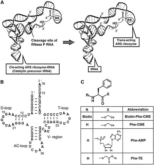
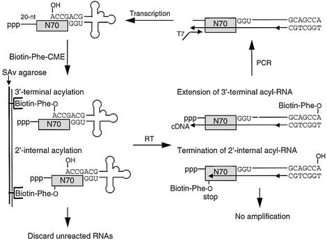
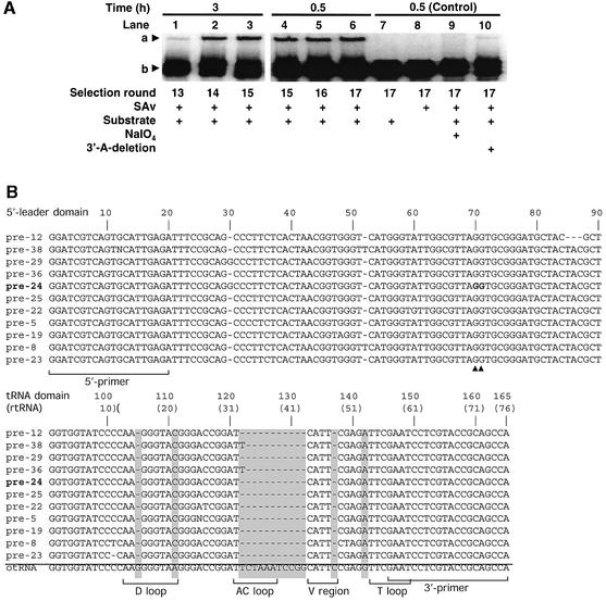
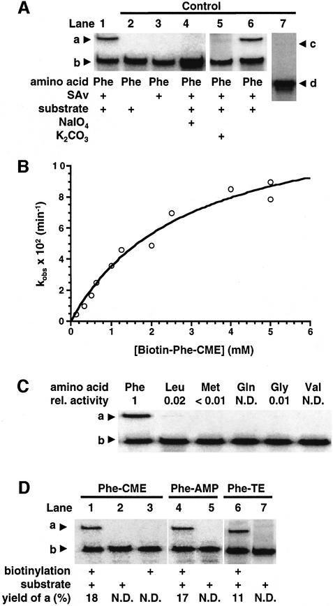
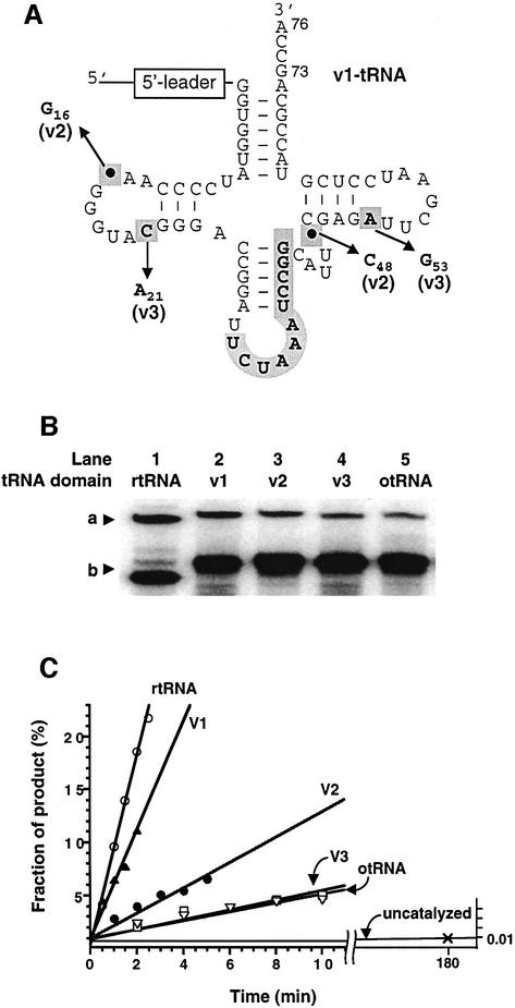
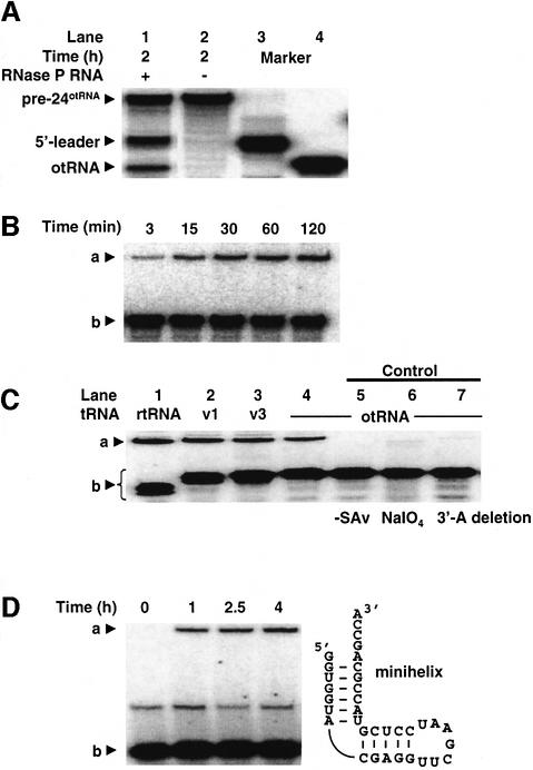
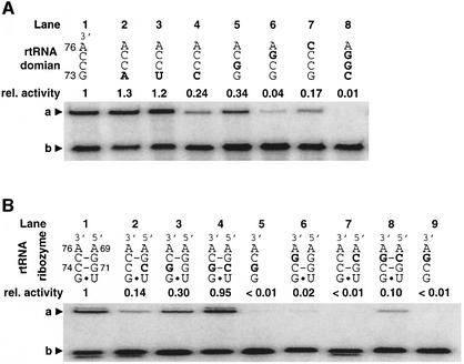
A set of catalysts for aminoacyl-tRNA synthesis is an essential component for translation. The RNA world hypothesis postulates that RNA catalysts could have played this role. Here we show an in vitro evolved precursor tRNA consisting of two domains, a catalytic 5'-leader sequence and an aminoacyl-acceptor tRNA. The 5'-leader sequence domain selectively self-charges phenylalanine on the 3'-terminus of the tRNA domain. This cis-acting ribozyme is susceptible to RNase P RNA, generating the corresponding 5'-leader segment and the mature tRNA. Moreover, the 5'-leader segment is able to aminoacylate the mature tRNA in trans. Mutational studies have revealed that C(74) and C(75) at the tRNA aminoacyl-acceptor end form base pairs with G71 and G70 of the trans-acting ribozyme. Such Watson-Crick base pairing with tRNA has been observed in RNase P RNA and 23S rRNA, suggesting that all three ribozymes use a similar mechanism for the recognition of the aminoacyl-acceptor end. Our demonstrations indicate that catalytic precursor tRNAs could have provided the foundations for the genetic coding system in the proto-translation system.


Figures

None
Fig. 1. Schematic representation of a catalytic pre-tRNA. (A) Catalytic pre-tRNA with self-aminoacylation activity (left). The amino acid substrate (the amino acid side chain and leaving group are shown with aa and X, respectively) binds to the 5′-leader ribozyme domain, and the nucleophilic attack of the tRNA 3′-hydroxyl (indicated by a curved arrow) is accelerated. The cleavage site of RNase P RNA is shown by the straight arrow. Trans-acting ribozyme that aminoacylates the 3′-end of tRNA (right). (B) Secondary structure of artificial orthogonal suppressor tRNA (otRNA) used for the construction of the RNA pool. The base numbers of otRNA are assigned according to the tRNA numbering rule (Sprinzl et al., 1998). The abbreviations for tRNA loops are: AC, anticodon; V, variable; T, TΨC. (C) Chemical structure of the phenylalanyl substrates.
None
Fig. 2. Schematic representation of in vitro selection for self-aminoacylating pre-tRNAs. Abbreviations: N70, a random region with the length of 70 nt; SAv, streptavidin; RT, reverse transcription; cDNA, complementary DNA; T7, T7 RNA promoter.
None
Fig. 3. Selection results. (A) Selection of catalytic pre-tRNAs. Autoradiogram showing self-aminoacylation activity as a function of selection cycle. One micromolar RNA and 1 mM Biotin-Phe-CME were used for the reaction in each selection round. a, Biotin-Phe-RNA complexed with SAv; b, RNA pool of each selection round. For periodate oxidation (lane 9), round 17 RNA was treated with 10 mM NaIO4 at 0°C for 1 h and ethanol precipitated prior to the aminoacylation reaction. Round 17 RNA lacking its 3′-A (lane 10) was prepared by transcription of a shortened template. (B) Sequence alignment of active clones isolated from round 17 RNA. In the tRNA domain, the observed deletions and mutations are highlighted by gray boxes, and this truncated tRNA domain sequence is referred to as rtRNA. For the alignment of the tRNA domain, the wild-type otRNA is shown together with the rtRNA sequences. The bases of the tRNA domain are numbered continuously from the 5′-leader domain sequence, and those of rtRNA in parentheses are numbered based on the rule of tRNA numbering (Sprinzl et al., 1998). G70 and G71 of the 5′-leader domain described in Figure 7B are shown by bold letters (their positions are indicated by triangles).
None
Fig. 4. Self-aminoacylation activity of pre-24. a, Biotin-aminoacyl-pre-24 complexed with SAv; b, pre-24; c, Biotin-Phe-otRNA complexed with SAv; d, otRNA. (A) Self-aminoacylation of pre-24. Reactions were carried out in the presence of 1 µM RNA and 1 mM Biotin-Phe-CME (lanes 1, 2, 4, 5 and 6), 5 mM Biotin-Phe-CME (lane 7) or in the absence of substrate (lane 3). For periodate oxidation (lane 4), pre-24 was treated with 10 mM NaIO4 at 0°C for 1 h and ethanol precipitated prior to the aminoacylation reaction. For mild base hydrolysis of the aminoacyl group on pre-24 (lane 5), Biotin-Phe-pre-24 RNA (lane 1) was treated with 50 mM K2CO3 for 15 min at 37°C. The deacylated pre-24 from lane 5 was recovered by ethanol precipitation, then used for aminoacylation under the same conditions as lane 1 (lane 6). The incubation time was 30 min (lanes 1–6) or 3 h (lane 7). (B) Michaelis–Menten-like plot of the initial rates observed in self-aminoacylation of pre-24. The observed rate at each substrate concentration was determined by a slope of the linear region in a plot of time versus fraction of product with five time points. Data were fitted to the Michaelis–Menten non-linear regression curve, giving the values described in the text. (C) Amino acid selectivity of pre-24. Abbreviations: rel. activity, relative catalytic activity based on Biotin-Phe-CME; N.D., not detectable. Reactions were carried out in the presence of 1 µM RNA and 1 mM Biotin-aa-CME at 25°C for 2 h. (D) Comparison of self-aminoacylation activity of pre-24 using three distinct amino acid esters. Reactions were performed in the presence of 0.5 µM pre-24 and 5 mM Phe-CME (lane 1) or Phe-TE (lane 6) at 25°C for 30 min or 5 mM Phe-AMP (lane 4) on ice for 30 min. After aminoacylation, the biotinylation was performed as described in Materials and methods. Control experiments in the absence of substrate (lane 3) or biotinylation (lanes 2, 5 and 7) were performed.
None
Fig. 5. Structure and self-aminoacylation activity of pre-24 and its variants. (A) Structure of otRNA variants introduced in the otRNA domain of pre-24. The mutations and deletions observed in rtRNA are highlighted by bold letters in gray boxes (see also Figure 1B). In order to distinguish the base numbering of tRNA from that of the 5′-leader domain, the subscript number is used for the assignment of tRNA bases according to the tRNA numbering rule (see Figure 3B). The structure of each variant is as follows: v1-tRNA (its structure is shown), in which the original anticodon region of otRNA was restored but the point mutations and deletions observed in rtRNA were maintained; v2-tRNA, in which the two point deletions were restored into v1-tRNA but the mutations were maintained; v3-tRNA, in which the two point mutations were restored into v1-tRNA but the deletions were maintained. (B) Comparison of self-aminoacylation activity of pre-24 and its variants with different tRNA domains. a, Biotin-Phe-pre-24 or variants complexed with SAv; b, pre-24 or variants. Reactions were carried out in the presence of 1 µM pre-24 (lane 1), variants (lanes 2–4) or pre-24otRNA (lane 5), incubating with 1 mM Biotin-Phe-CME at 25°C for 2 h. (C) Initial rates of self-aminoacylation of pre-24 and its variants. Reactions were carried out in the presence of 0.5 µM pre-24 (rtRNA) or its variants (v1–v3-tRNA and otRNA) incubated with 5 mM Biotin-Phe-CME at 25°C. The observed rate constants (kobs) are 0.089 min–1 (rtRNA), 0.051 min–1 (v1-tRNA), 0.012 min–1 (v2-tRNA), 0.0047 min–1 (v3-tRNA) and 0.0043 min–1 (otRNA).
None
Fig. 6. RNase P RNA cleavage of pre-24otRNA and the 5′-leader ribozyme-catalyzed tRNA aminoacylation in trans. (A) Cleavage of pre-24otRNA by RNase P RNA. The 32P-body-labeled pre-24otRNA was treated with RNase P RNA for 2 h, resulting in the cleavage of 23% of pre-24otRNA (lane 1). The absence of RNase P RNA yielded no cleaved product (lane 2). The marker RNAs (5′-leader segment in lane 3 and otRNA in lane 4) were prepared by in vitro transcription using the corresponding DNA segments. (BTrans-aminoacylation of otRNA. The autoradiogram displays the time course of 5′-leader ribozyme-catalyzed aminoacylation of 5′-32P-labeled otRNA. a, Biotin-Phe-otRNA complexed with SAv; b, otRNA. The RNase P-digested RNA fragments of pre-24otRNA (2 µM) were used for aminoacylation of 5′-32P-labeled otRNA (0.5 µM), giving kobs = 1.0 × 10⁻³ min–1. (CTrans-aminoacylation of tRNA variants. rtRNA, v1, v3 and otRNA are the fragment of the tRNA domain described in Figures 1B and 5A. a, Biotin-Phe-otRNA or tRNA variants complexed with SAv; b, otRNA or tRNA variants. tRNA variants were prepared by in vitro transcription using the corresponding DNA segments. Controls: lane 5, the absence of SAv; lane 6, otRNA treated with NaIO4 was used for aminoacylation; lane 7, otRNA lacking its 3′-A was used for aminoacylation. (DTrans-aminoacylation of a minihelix RNA. The autoradiogram depicts the time course of 5′-leader ribozyme-catalyzed aminoacylation of a minihelix RNA. a, Biotin-Phe-minihelix RNA complexed with SAv; b, minihelix RNA.
None
Fig. 7. Mutational studies of the aminoacyl-acceptor end of tRNA and 5′-leader ribozyme. The introduced mutations are highlighted by bold letters. The base numbers of 5′-leader ribozyme and rtRNA are assigned according to Figure 3B. (A) Self-aminoacylation activity of pre-24 and its mutants containing mutations at the acceptor end of the tRNA domain. Abbreviations: rel. activity, relative catalytic activity based on wild-type pre-24. a, Biotin-Phe-pre-24 or its mutants complexed with SAv; b, pre-24 or its mutants. The mutations were introduced into the pre-24 DNA template by PCR site-directed mutagenesis using the corresponding 3′-primer. Self-aminoacylation was carried out in the presence of 1 mM Biotin-Phe-CME and 1 µM RNA at 25°C for 30 min. (BTrans-aminoacylation activity and compensatory mutations of rtRNA and 5′-leader ribozyme. Abbreviations: rel. activity, relative catalytic activity based on the wild-type pair of rtRNA and 5′-leader ribozyme. a, Biotin-Phe-rtRNA or its mutant complexed with SAv; b, rtRNA or its mutant. Mutant rtRNA and 5′-leader ribozyme were independently transcribed in vitro, and the trans-aminoacylation was carried out in the presence of 1 µM mutant 5′-leader ribozyme and 0.5 µM mutant rtRNA at 25°C for 30 min.

References

    1. Arnez J.G. and Moras,D. (1997) Structural and functional considerations of the aminoacylation reaction. Trends Biochem. Sci., 22, 211–216. - PubMed
    1. Bartel D.P. and Szostak,J.W. (1993) Isolation of new ribozymes from a large pool of random sequences. Science, 261, 1411–1418. - PubMed
    1. Berg P. (1958) The chemical synthesis of amino acid adenylates. J. Biol. Chem., 253, 608–611. - PubMed
    1. Beuning P.J. and Musier-Forsyth,K. (1999) Transfer RNA recognition by aminoacyl-tRNA synthetases. Biopolymers, 52, 1–28. - PubMed
    1. Busch S., Kirsebom,L.A., Notbohm,H. and Hartmann,R.K. (2000) Differential role of the intermolecular base-pairs G292-C75 and G293-C74 in the reaction catalyzed by Escherichia coli RNase P RNA. J. Mol. Biol., 299, 941–951. - PubMed

Publication types