. 2001 Apr 2;153(1):63-74.
doi: 10.1083/jcb.153.1.63.

The Vfl1 Protein in Chlamydomonas localizes in a rotationally asymmetric pattern at the distal ends of the basal bodies

Affiliations

The Vfl1 Protein in Chlamydomonas localizes in a rotationally asymmetric pattern at the distal ends of the basal bodies

C D Silflow et al. J Cell Biol. .

Abstract

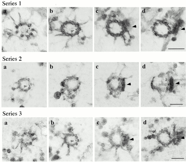
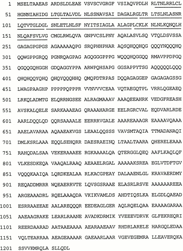
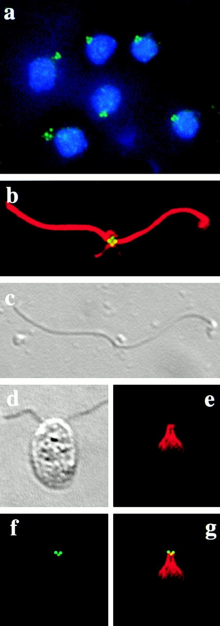
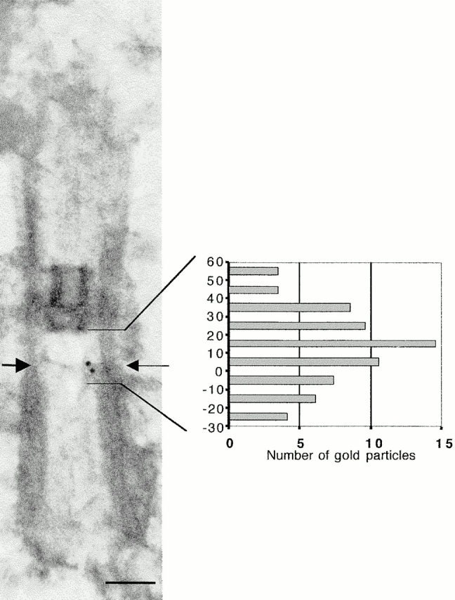
In the unicellular alga Chlamydomonas, two anterior flagella are positioned with 180 degrees rotational symmetry, such that the flagella beat with the effective strokes in opposite directions (Hoops, H.J., and G.B. Witman. 1983. J. Cell Biol. 97:902-908). The vfl1 mutation results in variable numbers and positioning of flagella and basal bodies (Adams, G.M.W., R.L. Wright, and J.W. Jarvik. 1985. J. Cell Biol. 100:955-964). Using a tagged allele, we cloned the VFL1 gene that encodes a protein of 128 kD with five leucine-rich repeat sequences near the NH(2) terminus and a large alpha-helical-coiled coil domain at the COOH terminus. An epitope-tagged gene construct rescued the mutant phenotype and expressed a tagged protein (Vfl1p) that copurified with basal body flagellar apparatuses. Immunofluorescence experiments showed that Vfl1p localized with basal bodies and probasal bodies. Immunogold labeling localized Vfl1p inside the lumen of the basal body at the distal end. Distribution of gold particles was rotationally asymmetric, with most particles located near the doublet microtubules that face the opposite basal body. The mutant phenotype, together with the localization results, suggest that Vfl1p plays a role in establishing the correct rotational orientation of basal bodies. Vfl1p is the first reported molecular marker of the rotational asymmetry inherent to basal bodies.


Figures

Figure 9
Figure 9
Rotationally asymmetric localization of Vfl1p. Three series of adjacent serial sections (50-nm sections) of isolated NFAps labeled by the immunogold preembedding method show the positions of the gold particles with respect to doublet microtubules 9, 1, and 2. The images are viewed from the distal end of the basal body. Numbers 9, 1, and 2 identify these triplets based on their association with the distal striated fiber. Gold particles are marked by light arrowheads; the distal striated fiber is marked by a dark arrowhead. In series 1 a, the upper gold particle is nearest to doublet 2 and the lower particle is nearest to doublet 1. In series 1 b, the particle is nearest to doublet 1. In series 2 a, the gold particle is nearest to doublet 1. In series 3 b, the particle is nearest to doublet 1. Bars, 0.2 μm.
Figure 1
Figure 1
Genomic region of the VFL1 gene. The top bar represents a 16-kb fragment of genomic DNA cloned in the plasmid pW6 that rescues the phenotype of the vfl1-2 mutation when transformed into mutant cells. SstI sites are indicated. Subclones of the genomic region (represented by solid black bars) were tested for their ability to rescue the mutation. A 9.2-kb region within the complementing subclone pW6-10.0 was sequenced. GeneMark analysis of the sequence and RT-PCR confirmation of the predicted gene structure identified a coding region spanning 7.8 kb. In DNA from the vfl1-2 allele, a deletion of ∼4 kb was mapped to the region indicated by the bottom hatched bar.
Figure 2
Figure 2
The VFL1 gene encodes a transcript of ∼4.0 kb. Polyadenylated RNA (10 μg/lane) was electrophoresed on a denaturing gel and blotted to a nylon membrane. The RNA was hybridized with a labeled probe consisting of the first exon of the gene. RNA was obtained from: lane 1, wild-type cells; lane 2, vfl1-1 cells; and lane 3, vfl1-2 cells. The lower panel is an RNA loading control showing hybridization with a radioactive probe for the CRY1 gene (Nelson et al. 1994).
Figure 3
Figure 3
Predicted sequence of the Vfl1 protein. The five LRRs are underlined.
Figure 4
Figure 4
Summary of the predicted Vfl1 protein structure. (a) The LRR region (vertical bars) is located near the NH2 terminus, and the coiled coil regions (hatched bars) are at the COOH terminus. The position where the amino acid sequence is disrupted in the protein encoded by the vfl1-1 allele is shown by a vertical open arrow. The five LRR sequences are aligned, with the consensus sequence shown below. (b) COILS output for the Vfl1p using a window size of 21 (Lupas et al. 1991).
Figure 5
Figure 5
The Vfl1 protein copurifies with the basal body apparatus. Whole cell proteins (lanes 1 and 3) and proteins from NFAps (lanes 2 and 4) were isolated from VFL1-2-R2 cells expressing the VFL1 HA-tagged gene construct (lanes 1 and 2) and from VFL1-2-R29 cells expressing an untagged VFL1 gene construct (lanes 3 and 4). The proteins were separated by SDS-PAGE and blotted to nylon membrane. (a) The anti-HA high affinity antibody was used to identify the tagged Vfl1p which migrates as a 140,000-M r protein. (b) The blot was stripped and reprobed with an anti–β-tubulin antibody as a control. Silver-stained lanes from the gel are shown in panel c.
Figure 6
Figure 6
Immunofluorescence microscopy of NFAps and whole cells. (a) NFAps isolated from cells expressing HA-tagged Vfl1p were stained with rat anti-HA antibody, followed by FITC-conjugated goat anti–rat IgG (green). The DNA was labeled with DAPI (blue). (b) A single apparatus was costained with rat anti-HA and rabbit anti–α-tubulin antibodies, followed by FITC-conjugated goat anti–rat (green) and Texas red–conjugated goat anti–rabbit (red) IgGs. (c) DIC microscopy of the apparatus labeled in panel b. The nucleus is missing in this apparatus. (d) DIC microscopy of an intact cell expressing HA-tagged Vfl1p, analyzed by immunofluorescence microscopy (e–g). (e) Texas red channel showing the centrin distribution with the distal striated fiber that connects the two basal bodies. (f) FITC channel showing Vfl1p localization with two intensely stained dots and one weakly stained dot. (g) Superimposed images from panels e and f.
Figure 7
Figure 7
Ultrastructural localization of Vfl1p by immunogold preembedded labeling of isolated NFAps. (a and b) Longitudinal sections of basal bodies (95-nm sections). Arrows indicate the point at the distal end of the basal body where the C tubules end and the doublet microtubules of the axoneme begin. The electron-dense H-shaped structure is the transition region. Dark arrowheads indicate the distal striated fibers. Gold particles are indicated by light arrow heads. (c and d) Cross-sections made at the distal ends of basal bodies are viewed from the proximal end of the basal body (95-nm sections). Arrows indicate transitional fibers connecting the nine triplet microtubules with the plasma membrane. Gold particles are located within the basal body cylinder. (e and f) Cross-sections of the proximal region of the apparatus showing basal body pairs and probasal bodies. Gold particles indicated by arrowheads are found in positions expected for probasal bodies. e, 200-nm section; f, 50-nm section. Bars, 0.2 μm.
Figure 8
Figure 8
Localization of Vfl1p relative to the distal ends of the triplet microtubules. The positions of 67 gold particles on 49 longitudinal basal body sections were determined with respect to the end of the triplet microtubules of the basal body (arrow). The data were normalized as described in Materials and Methods and plotted to indicate the distribution distal (positive numbers) or proximal (negative numbers) to the end of the triplet microtubules. Bar, 0.2 μm

References

    1. Adams G.M.W., Wright R.L., Jarvik J.W. Defective temporal and spatial control of flagellar assembly in a mutant of Chlamydomonas reinhardtii with variable flagellar number. J. Cell Biol. 1985;100:955–964. - PMC - PubMed
    1. Afzelius B. Electron microscopy of the sperm tail. J. Biophys. Biochem. Cytol. 1959;2:269–278. - PMC - PubMed
    1. Allen R.D. The morphogenesis of basal bodies and accessory structures of the cortex of the ciliated protozoan Tetrahymena pyriformis . J. Cell Biol. 1969;40:716–733. - PMC - PubMed
    1. Andersen S.S. Molecular characteristics of the centrosome. Int. Rev. Cytol. 1999;187:51–109. - PubMed
    1. Anstrom J.A. Organization of the ciliary basal apparatus in embryonic cells of the sea urchin, Lytechinus pictus . Cell Tissue Res. 1992;269:305–313. - PubMed

Publication types

Associated data