. 2001 Apr 2;153(1):149-58.
doi: 10.1083/jcb.153.1.149.

Regulation of Op18 during spindle assembly in Xenopus egg extracts

Affiliations

Regulation of Op18 during spindle assembly in Xenopus egg extracts

P P Budde et al. J Cell Biol. .

Abstract

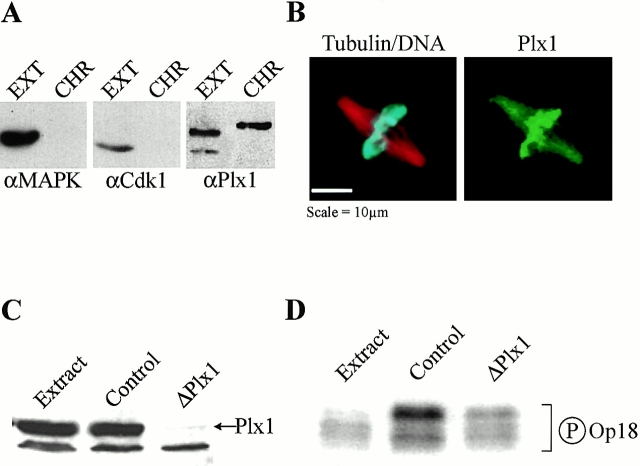
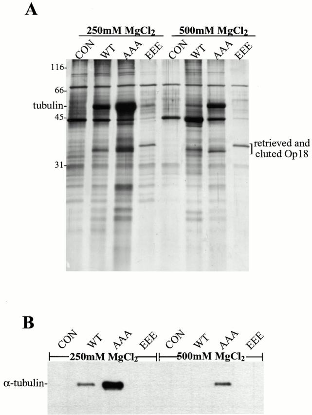
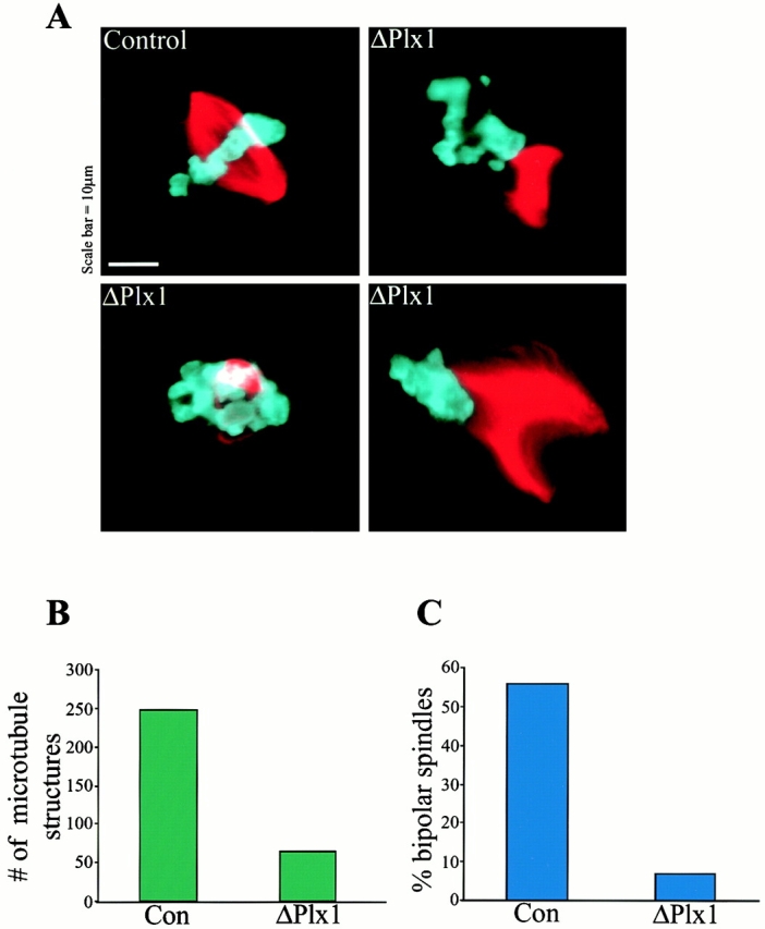
Oncoprotein 18 (Op18) is a microtubule-destabilizing protein that is negatively regulated by phosphorylation. To evaluate the role of the three Op18 phosphorylation sites in Xenopus (Ser 16, 25, and 39), we added wild-type Op18, a nonphosphorylatable triple Ser to Ala mutant (Op18-AAA), and to mimic phosphorylation, a triple Ser to Glu mutant (Op18-EEE) to egg extracts and monitored spindle assembly. Op18-AAA dramatically decreased microtubule length and density, while Op18-EEE did not significantly affect spindle microtubules. Affinity chromatography with these proteins revealed that the microtubule-destabilizing activity correlated with the ability of Op18 to bind tubulin. Since hyperphosphorylation of Op18 is observed upon addition of mitotic chromatin to extracts, we reasoned that chromatin-associated proteins might play a role in Op18 regulation. We have performed a preliminary characterization of the chromatin proteins recruited to DNA beads, and identified the Xenopus polo-like kinase Plx1 as a chromatin-associated kinase that regulates Op18 phosphorylation. Depletion of Plx1 inhibits chromatin-induced Op18 hyperphosphorylation and spindle assembly in extracts. Therefore, Plx1 may promote microtubule stabilization and spindle assembly by inhibiting Op18.


Figures

Figure 5
Figure 5
Plx1 is a chromatin-associated kinase and its depletion inhibits chromatin-induced Op18 hyperphosphorylation. (A) Immunoblot analysis of total extract (EXT) and proteins eluted from mitotic chromatin beads using 0.5 M KCl (CHR) probed with antibodies against MAP kinase, cdk1, and Plx1. (B) Immunofluorescence micrograph showing Plx1 localization to chromatin and microtubules in sperm spindles assembled in Xenopus egg extracts. Microtubules appear red, sperm chromosomes blue, and Plx1 green. (C) Immunoblot of total extract (Extract), IgG-depleted (Control), and Plx1-depleted (ΔPlx1) extracts shows that >99% of Plx1 has been removed. (D) Autoradiogram showing [γ32P]phosphate incorporation in Op18 immunoprecipitated from mitotic extract (Extract) or in IgG-depleted (Control) or Plx1-depleted (ΔPlx1) mitotic extract + chromatin beads.
Figure 1
Figure 1
Differential effects of phosphorylation site mutants of Op18 on spindle assembly reactions in Xenopus egg extracts. (A) Effects of 6 μM Op18-WT, AAA (all three Serine phosphorylation sites mutated to Alanine) or EEE (all three Serine phosphorylation sites mutated to Glutamic acid) on spindle assembly in Xenopus egg extracts. Microtubules appear red and sperm chromosomes blue. (B) Biochemical quantification of depolymerizing activities shown by antitubulin immunoblot of microtubules pelleted from extracts in the presence of 6 or 12 μM Op18-WT, -AAA, and -EEE. The control (CON) is buffer addition in both A and B.
Figure 2
Figure 2
Nonphosphorylatable Op18 binds tightly to tubulin in extracts. (A) Silver-stained gel of MgCl2 elutions of control, ZZ-WT, -AAA, and -EEE Op18 bound to IgG Sepharose retrieved from extracts. (B) The elutions from A were blotted for α tubulin. The control (CON) is buffer addition in both A and B.
Figure 3
Figure 3
Chromatin assembles on DNA beads. (A) Silver stained gel of proteins associated with DNA beads and control beads in interphase (I) and mitotic (M) extracts. Arrowheads point to bands that are different between I and M. Histones (H) are indicated. (B) Western blot of the same samples from A probed with antibodies to Xklp1 and Topoisomerase II. (C) Micrococcal nuclease digestion of DNA beads for 1, 2, or 5 min; mononucleosomes (mono) and dinucleosomes (di) are indicated. Outer lanes are 123-bp markers.
Figure 4
Figure 4
Chromatin-associated proteins phosphorylate recombinant Op18 in vitro. (A) Autoradiogram of recombinant Op18 showing [γ32P]phosphate incorporation after incubation with interphase or mitotic extract, or chromatin beads isolated from interphase or mitotic extract. (B) Quantification of in vitro Op18 kinase data comparing total extract (I, M extract) and 0.5 M KCl-eluted chromatin proteins (I, M eluate).
Figure 6
Figure 6
Plx1 depletion causes defects in microtubule polymerization and spindle assembly. (A) Representative microtubule structures found in control- and Plx1-depleted extracts. (B) Quantification of spindle defects showing the decrease in total microtubule structures formed in Plx1-depleted (ΔPlx1, 64) compared with IgG-depleted (Con, 247), and (C) the small percentage of normal bipolar spindles formed in Plx1-depleted extract. A total of 933 structures were counted in three separate experiments.
Figure 7
Figure 7
Model for Op18 regulation of microtubule dynamics in Xenopus. The curved line represents Op18 and P is a phosphate group. Unphosphorylated Op18 promotes microtubule depolymerization by sequestering tubulin. Op18 that encounters mitotic chromatin is phosphorylated by Plx1, preventing the Op18-tubulin interaction and promoting microtubule stabilization.

References

    1. Andersen S. Spindle assembly and the art of regulating microtubule dynamics by MAPs and Stathmin/Op18. Trends Cell Biol. 2000;10:261–267. - PubMed
    1. Andersen S.S.L., Ashford A.J., Tournebize R., Gavet O., Sobel A., Hyman A.A., Karsenti E. Mitotic chromatin regulates phosphorylation of Stathmin/Op18. Nature. 1997;389:640–643. - PubMed
    1. Arnaud L., Pines J., Nigg E.A. GFP tagging reveals human Polo-like kinase 1 at the kinetochore/centromere region of mitotic chromosomes. Chromosoma. 1998;107:424–429. - PubMed
    1. Bahler J., Steever A.B., Wheatley S., Wang Y.-L., Pringle J.R., Gould K.L., McCollum D. Role of polo kinase and Mid1p in determining the site of cell division in fission yeast. J. Cell Biol. 1998;143:1603–1616. - PMC - PubMed
    1. Belmont L.D., Mitchison T.J. Identification of a protein that interacts with tubulin dimers and increases the catastrophe rate of microtubules. Cell. 1996;84:623–631. - PubMed

Publication types