. 2001 Apr 2;153(1):159-68.
doi: 10.1083/jcb.153.1.159.

The surveillance mechanism of the spindle position checkpoint in yeast

Affiliations

The surveillance mechanism of the spindle position checkpoint in yeast

N R Adames et al. J Cell Biol. .

Abstract

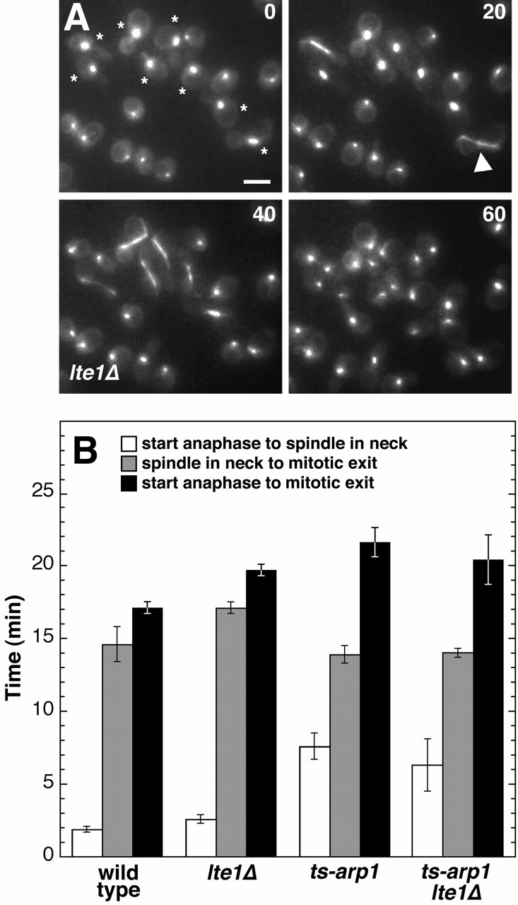
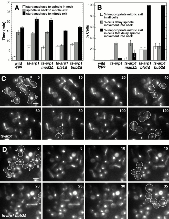
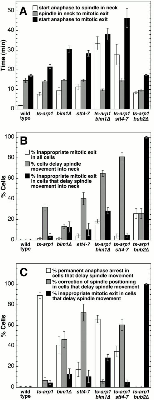
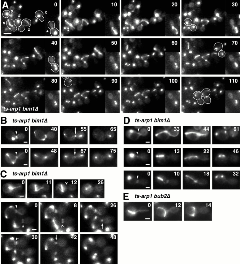
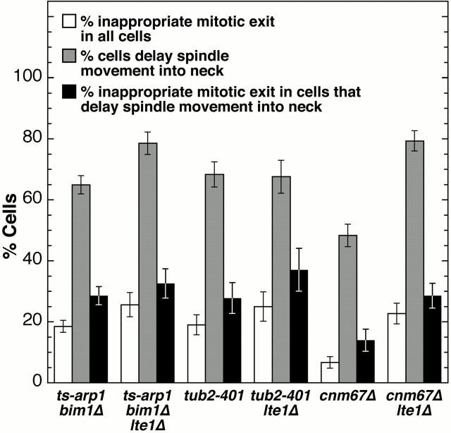
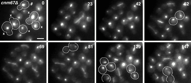
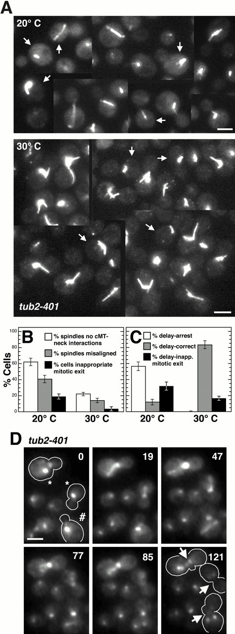
The spindle position checkpoint in Saccharomyces cerevisiae delays mitotic exit until the spindle has moved into the mother-bud neck, ensuring that each daughter cell inherits a nucleus. The small G protein Tem1p is critical in promoting mitotic exit and is concentrated at the spindle pole destined for the bud. The presumed nucleotide exchange factor for Tem1p, Lte1p, is concentrated in the bud. These findings suggested the hypothesis that movement of the spindle pole through the neck allows Tem1p to interact with Lte1p, promoting GTP loading of Tem1p and mitotic exit. However, we report that deletion of LTE1 had little effect on the timing of mitotic exit. We also examined several mutants in which some cells inappropriately exit mitosis even though the spindle is within the mother. In some of these cells, the spindle pole body did not interact with the bud or the neck before mitotic exit. Thus, some alternative mechanism must exist to coordinate mitotic exit with spindle position. In both wild-type and mutant cells, mitotic exit was preceded by loss of cytoplasmic microtubules from the neck. Thus, the spindle position checkpoint may monitor such interactions.


Figures

Figure 1
Figure 1
Lte1p is not necessary for mitotic exit. (A) Still images from Video 1. Representative field showing mitosis in lte1Δ cells. Cells expressed GFP–tubulin to allow visualization of the spindle (bright thick bar of fluorescence) and cytoplasmic microtubules. Cells also expressed Hof1–GFP to allow visualization of mitotic exit. Hof1–GFP initially localizes to the septin ring structures at the neck (arrowhead). During mitotic exit, Hof1–GFP moves to the constricting actomyosin ring and then disappears from the neck (Lippincott and Li 1998a). Not all cells in the population expressed Hof1–GFP to the same extent. Time (min) is indicated in the upper right. (B) Loss of Lte1p has little effect on the kinetics of mitotic exit. Shown are the mean time intervals from the start of anaphase (spindle elongation) to spindle movement into the neck (white columns), from spindle movement into the neck to mitotic exit (spindle break down and constriction of the actomyosin ring; gray columns), and the total time for anaphase (black columns). The lte1Δ mutant was slightly slower than the wild-type strain (P < 0.01). Number of events for each column from left to right on the chart: wild type, 108, 83, 83; lte1Δ, 115, 112, 112; ts-arp1, 120, 116, 114; ts-arp1 lte1Δ, 48, 66, 48. Error bars represent SEM. Supplemental videos are available at http://www.jcb.org/cgi/content/full/153/1/159/DC1. Bar, 5 μm.
Figure 2
Figure 2
The spindle position checkpoint is completely abolished in bub2Δ and bfa1Δ mutants, but not in mad2Δ mutants. (A) Cytokinesis was delayed in ts-arp1 and ts-arp1 mad2Δ cells, but not in the ts-arp1 bfa1Δ and ts-arp1 bub2Δ mutants. Shown are the mean time intervals from the start of anaphase to spindle movement into the neck (white columns), from spindle movement into the neck to mitotic exit (gray columns), and the total time for anaphase (black columns). In the bfa1Δ and bub2Δ mutants, mitotic exit occurred in about half the time (P < 0.001), after spindle movement into the neck, compared with the wild-type strain. Number of events for each column from left to right on the chart: wild type, 108, 83, 83; ts-arp1, 120, 116, 114; ts-arp1 mad2Δ, 65, 71, 64; ts-arp1 bfa1Δ, 94, 91, 114; ts-arp1 bub2Δ, 68, 66, 92. Error bars represent SEM. (B) In the ts-arp1 and ts-arp1 mad2Δ strains, only a small fraction of cells that delayed spindle movement into the neck exited mitosis inappropriately. In the ts-arp1 bfa1Δ and ts-arp1 bub2Δ strains, all cells that delayed spindle movement into the neck underwent inappropriate mitotic exit. Comparison of the percentage of cells that underwent inappropriate mitotic exit (white columns) to the percentage of cells that delayed spindle movement into the neck by >20 min (gray columns) is shown. The frequency of inappropriate mitotic exit in cells that delayed spindle movement is also shown (black columns). Cell numbers: wild type, 217; ts-arp1, 287; ts-arp1 mad2Δ, 95; ts-arp1 bfa1Δ, 117; ts-arp1 bub2Δ, 93. Error bars represent standard error of the proportion (SEP). (C) Still images from Video 2. Mitosis in representative cells of a ts-arp1 mutant. The cells of interest are outlined in the first frame and again after they divide, or they are outlined in the last frame. Some cells (x) move the spindle into the neck within the normal time for mitotic exit. Other cells (y) delay spindle movement into the neck beyond the normal time for mitotic exit, but then correct the spindle position and divide. A third type of cell (z) does not move the spindle into the neck or exit mitosis during the course of the movie. Time (in min) is indicated. (D) Still images from Video 3. Mitosis in representative cells of a ts-arp1 bub2Δ mutant. Most cells (x) move the spindle into the neck within the normal time for mitotic exit. Those cells that do not move the spindle into the neck in time (*), exit mitosis at the normal time producing a binucleate mother and an anucleate daughter. Time (in min) is indicated. Supplemental videos are available at http://www.jcb.org/cgi/content/full/153/1/159/DC1. Bars, 5 μm.
Figure 3
Figure 3
Inappropriate mitotic exit in bim1Δ and stt4-7 mutants. (A) Exit from mitosis was delayed in the ts-arp1 bim1Δ and ts-arp1 stt4-7 mutants. Shown are the mean time intervals from the start of anaphase to spindle movement into the neck (white columns), from spindle movement into the neck to mitotic exit (gray columns), and the total time for anaphase (black columns). Notice the near lack of variability in the time interval from spindle movement into the neck to cytokinesis. Number of events for each column from left to right on the chart: wild type, 108, 83, 83; ts-arp1, 120, 116, 114; bim1Δ, 129, 170, 150; stt4-7, 49, 49, 55; ts-arp1 bim1Δ, 147, 135, 127; ts-arp1 stt4-7, 27, 33, 29; ts-arp1 bub2Δ, 68, 66, 92. Error bars represent SEM. (B) In the ts-arp1 bim1Δ mutant, a large fraction of cells that delayed spindle movement into the neck exited mitosis inappropriately. However, inappropriate mitotic exit did not occur in all ts-arp1 bim1Δ cells that delayed spindle movement into the neck. Comparison of the percentage of cells that underwent inappropriate mitotic exit (white columns) with the percentage of cells that delayed spindle movement into the neck by >20 min (gray columns) is shown. The frequency of inappropriate mitotic exit in cells that delayed spindle movement is also shown (black columns). Cell numbers: wild type, 217; ts-arp1, 287; bim1Δ, 290; stt4-7, 72; ts-arp1 bim1Δ, 259; ts-arp1 stt4-7, 100; ts-arp1 bub2Δ, 93. (C) Fate of cells that delay spindle movement into the neck. In the ts-arp1 mutant, most cells that delay spindle movement into the neck fail to move the spindle into the neck during the course of observation (white column). In the ts-arp1 stt4-7 mutant, many cells delay spindle movement into the neck but then correct the spindle position and exit mitosis (gray column). In the ts-arp1 bim1Δ mutant, many cells exit mitosis inappropriately (black column) instead of correcting the spindle position. Cell numbers: wild type, 0; ts-arp1, 92; bim1Δ, 39; stt4-7, 29; ts-arp1 bim1Δ, 168; ts-arp1 stt4-7, 81; ts-arp1 bub2Δ, 24. Error bars represent SEP.
Figure 4
Figure 4
Events that precede inappropriate mitotic exit in ts-arp1 bim1Δ cells. (A) Still images from Video 4. A representative field of ts-arp1 bim1Δ cells. The cells of interest are outlined in the first frame and again after they exit mitosis or in the last frame. Some cells (x) move the spindle into the neck within the normal time for mitotic exit. Other cells (y) delay spindle movement into the neck beyond the normal time for mitotic exit, but then correct the spindle position and divide. A third type of cell (z) does not move the spindle into the neck or exit mitosis during the course of the movie. Inappropriate mitotic exit occurs after abortive spindle movements into the neck (*; cell in inset taken from a different field in the same movie) or when cytoplasmic microtubules are lost from the neck (#). (B) Still images from Videos 5 and 6. In some ts-arp1 bim1Δ cells, spindle movement into the neck is severely delayed and so is exit from mitosis, as indicated by maintenance of Hof1–GFP on the septin ring (arrowhead). Once the spindle moves into the neck, mitotic exit and spindle disassembly proceed (arrow). (C) Still images from Videos 7 and 8. In ts-arp1 bim1Δ cells, the spindle delays movement into the neck and Hof1–GFP remains at the septin rings (arrowheads). Inappropriate mitotic exit occurs in the absence of SPB–neck interactions. Instead, cytokinesis and spindle disassembly (arrow) occur after the loss of cytoplasmic microtubules from the neck (indented arrowhead). In the bottom cell of the lower series, cytoplasmic microtubules fail to penetrate or touch the neck at any time during anaphase. (D) Still images from Videos 9–11. Examples of ts-arp1 bim1Δ cells, in which inappropriate mitotic exit occurred after the spindle moved toward and touched the neck without completely penetrating the neck. Spindle disassembly and cytokinesis occurred while the SPB was at the neck and often resulted in the shearing of a microtubule within the bud. The Hof1–GFP disappears from the neck after spindle disassembly. (E) Still images from Video 12. In ts-arp1 bub2Δ cells, inappropriate mitotic exit occurred without SPB–neck interactions or loss of cytoplasmic microtubules from the neck. Times are indicated in the upper right. Supplemental videos are available at http://www.jcb.org/cgi/content/full/153/1/159/DC1. Bars: (A) 5 μm; (B–E) 2 μm.
Figure 5
Figure 5
Lte1p is not required for inappropriate mitotic exit. Deletion of LTE1 does not decrease the frequency of inappropriate mitotic exit in mutants affecting cytoplasmic microtubule function—bim1Δ, tub2-401, and cnm67Δ. Shown are the percentage of cells that exit mitosis inappropriately (white columns), the percentage of cells that delay spindle movement into the neck (gray columns), and the percentage of cells that delay spindle movement into the neck and exit mitosis inappropriately (black columns). Cell numbers for each column from left to right: ts-arp1 bim1Δ, 259, 259, 168; ts-arp1 bim1Δ lte1Δ, 121, 121, 95; tub2-401, 142, 142, 97; tub2-401 lte1Δ, 80, 80, 54; cnm67Δ, 178, 178, 86; cnm67Δ lte1Δ, 150, 150, 119. Error bars represent SEP.
Figure 7
Figure 7
Inappropriate mitotic exit in cnm67Δ cells. Still images from Video 14. Representative cells showing two examples of inappropriate mitotic exit, one after the SPB touches the neck (*), and one without any SPB–neck interactions (#). Also shown are cells that move the spindle into the neck within the normal time for mitotic exit (x). Cells of interest are outlined. Supplemental videos are available at http://www.jcb.org/cgi/content/full/153/1/159/DC1. Bar, 5 μm.
Figure 6
Figure 6
Loss of cytoplasmic microtubules and inappropriate mitotic exit in tub2-401 cells. (A) At semipermissive temperatures, cytoplasmic microtubule staining is greatly reduced. At 20°C, many of the spindles are misaligned and lack cytoplasmic microtubules or show no interactions between short cytoplasmic microtubules and the neck (arrows). Misaligned spindles lacking cytoplasmic microtubule interactions with the neck are also present at 30°C but are much less frequent. Shown are collages of representative cells. (B) Frequency of the lack of cytoplasmic microtubule interactions with the neck (white columns), spindle misalignment (gray columns), and inappropriate mitotic exit (black columns) in tub2-401 cells. Cell numbers for each column from left to right: 20°C, 108, 108, 142; 30°C, 150, 150, 54. (C) The spindle position checkpoint is intact in the tub2-401 mutant. At 20°C, the majority of cells that delay spindle movement into the neck by >40 min arrest with an anaphase spindle at the neck on the mother side of the neck (white columns). At the lower temperature, few cells that delay spindle movement into the neck correct the defect and move the spindle into the neck (gray columns). Many of these cells undergo inappropriate mitotic exit (black columns). Cell numbers: 20°C, 85; 30°C, 12. (D) Still images from Video 13. Events preceding inappropriate mitotic exit in the tub2-401 mutant. Cells of interest are outlined for clarity. In this series, two cells exit mitosis inappropriately after an SPB touches the neck (*). One cell exits mitosis without any SPB–neck interaction (#). Note the poor alignment of the spindle with respect to the mother–bud axis in this cell. After mitotic exit, Hof1–GFP disappears or immediately relocates to the necks of the new buds (arrows). Error bars represent SEP. Supplemental videos are available at http://www.jcb.org/cgi/content/full/153/1/159/DC1. Bars, 5 μm.

References

    1. Adames N.R., Cooper J.A. Microtubule interactions with the cell cortex causing nuclear movements in Saccharomyces cerevisiae . J. Cell Biol. 2000;149:863–874. - PMC - PubMed
    1. Amatruda J.F., Cooper J.A. Purification, characterization and immunofluorescence localization of Saccharomyces cerevisiae capping protein. J. Cell Biol. 1992;117:1067–1076. - PMC - PubMed
    1. Bardin A.J., Visintin R., Amon A. A mechanism for coupling exit from mitosis to partitioning of the nucleus. Cell. 2000;102:21–31. - PubMed
    1. Bi E., Maddox P., Lew D.J., Salmon E.D., McMillan J.N., Yeh E., Pringle J.R. Involvement of an actomyosin contractile ring in Saccharomyces cerevisiae cytokinesis. J. Cell Biol. 1998;142:1301–1312. - PMC - PubMed
    1. Bloecher A., Venturi G.M., Tatchell K. Anaphase spindle position is monitored by the BUB2 checkpoint. Nat. Cell Biol. 2000;2:556–558. - PubMed

Publication types

MeSH terms