. 2001 Apr 2;153(1):169-76.
doi: 10.1083/jcb.153.1.169.

Nucleolar components involved in ribosome biogenesis cycle between the nucleolus and nucleoplasm in interphase cells

Affiliations

Nucleolar components involved in ribosome biogenesis cycle between the nucleolus and nucleoplasm in interphase cells

D Chen et al. J Cell Biol. .

Abstract

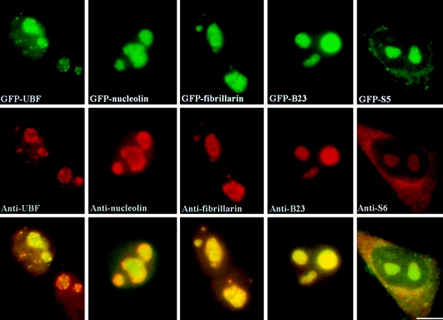
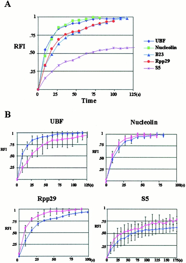
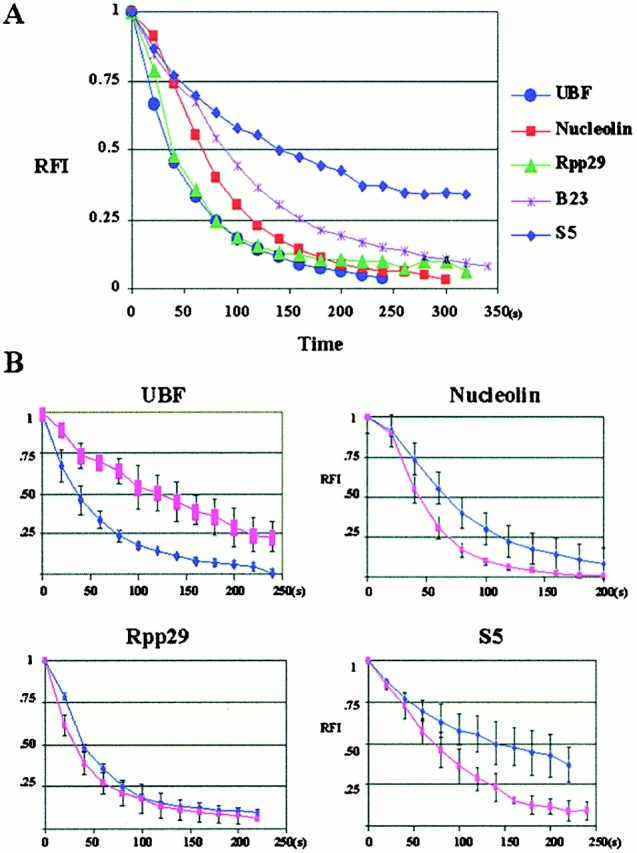
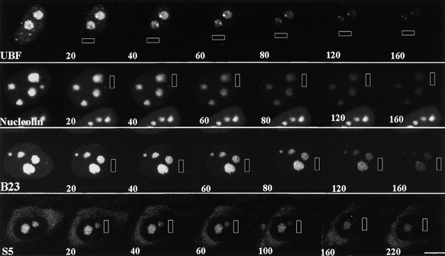
We examined the mobilities of nucleolar components that act at various steps of the ribosome biogenesis pathway. Fluorescence recovery after photobleaching (FRAP) and fluorescence loss in photobleaching (FLIP) analyses demonstrate that factors involved in rRNA transcription (upstream-binding factor [UBF]), processing (nucleolin, fibrillarin, and RNase MRP subunits, Rpp29), and ribosome assembly (B23) exchange rapidly between the nucleoplasm and nucleolus. In contrast, the mobilities of ribosomal subunit proteins (S5, L9) are much slower. Selective inhibition of RNA polymerase I transcription does not prevent the exchanges but influences the rates of exchange differentially for different nucleolar components. These findings suggest that the rapid exchange of nucleolar components between the nucleolus and nucleoplasm may represent a new level of regulation for rRNA synthesis. The different dynamic properties of proteins involved in different steps of ribosome biogenesis imply that the nucleolar association of these proteins is due to their specific functional roles rather than simply their specific nucleolar-targeting events.


Figures

Figure 1
Figure 1
GFP fusion proteins and their corresponding endogenous proteins localize to the same subcellular regions. Top panels show the localization of GFP fusion proteins as indicated. The middle panels show the localization of the endogenous proteins immunolabeled with specific antibodies. Lower panels show the mergers of the corresponding red and green panels. Bar, 10 μm.
Figure 2
Figure 2
FRAP analyses demonstrate that the examined nucleolar components exchange rapidly between the nucleolus and nucleoplasm. Arrowheads indicate the sites of bleaching and numbers represent the time (s) after photobleaching. BL is the first image obtained immediately after photobleaching. Bar, 10 μm.
Figure 3
Figure 3
Quantitative analyses of FRAP demonstrate that the FRAP rate of GFP ribosomal proteins is significantly slower than UBF and some of the factors involved in pre-rRNA processing. x, time after the photobleaching (s); y, relative fluorescence intensity in the testing nucleolus. (A) A summary of FRAP rates of various nucleolar components examined in this study. (B) Comparisons of the FRAP rates of some of the examined factors in cells treated or not treated with a low concentration of ActD that selectively inhibits pol I transcription. The dark blue line indicates the FRAP rate in untreated cells, and the pink line indicates the FRAP rate in treated cells.
Figure 6
Figure 6
Quantitative analyses of FLIP demonstrate that the GFP ribosomal proteins exit the nucleolus slower than UBF and some of the factors involved in pre-rRNA processing. x, cumulative duration of photobleaching (s); y, relative fluorescence intensity in the testing nucleolus. (A) A summary of FLIP rates of various nucleolar components examined in this study. (B) Comparisons of the FLIP rates of some of the examined factors in cells treated or not treated with a low concentration of ActD that selectively inhibits pol I transcription. The dark blue line indicates the FLIP rate in untreated cells, and the pink line indicates the FLIP rate in treated cells.
Figure 4
Figure 4
Quantitative analyses of FRAP demonstrate that the FRAP rate of all nucleolar GFP fusion proteins are similarly rapid in the nucleoplasm and are not affected during pol I transcription inhibition. x, time after the photobleaching (s); y, relative fluorescence intensity in the testing region. (A) A summary of FRAP rates of various nucleolar components examined in this study. (B) Comparisons of the FRAP rates of some of the examined factors in cells treated or not treated with a low concentration of ActD that selectively inhibits pol I transcription. The dark blue line indicates the FRAP rate in untreated cells, and the pink line indicates the FRAP rate in treated cells.
Figure 5
Figure 5
FLIP analyses demonstrate that the examined nucleolar components exit the interphase nucleolus rapidly. The rectangles indicate the areas of bleaching, and the numbers represent the cumulative bleaching duration (s). Bar, 10 μm.

References

    1. Busch H., Smetana K. Nucleoli of tumor cells. In: Busch H., editor. The Nucleolus. Academic Press; New York: 1970.
    1. Carmo-Fonseca M., Mendes-Soares L., Campos I. To be or not to be in the nucleolus. Nat. Cell Biol. 2000;2:E107–E112. - PubMed
    1. Chan E.K.L., Imai H., Hamel J.C., Tan E.M. Human autoantibody to RNA polymerase I transcription factor hUBF. Molecular identity of nucleolus organizer region autoantigen NOR-90 and ribosomal RNA transcription upstream binding factor. J. Exp. Med. 1991;174:1239–1244. - PMC - PubMed
    1. Chan Y.L., Wool I.G. The primary structure of rat ribosomal protein S6. J. Biol. Chem. 1988;263:2891–2896. - PubMed
    1. Dousset T., Wang C., Verheggen C., Chen D., Hernandez-Verdun D., Huang S. Initiation of nucleolar assembly is independent of RNA polymerase I transcription. Mol. Biol. Cell. 2000;11:2705–2717. - PMC - PubMed

Publication types

MeSH terms