. 2001 Apr;107(7):813-22.
doi: 10.1172/JCI10947.

A novel mouse model of lipotoxic cardiomyopathy

Affiliations

A novel mouse model of lipotoxic cardiomyopathy

H C Chiu et al. J Clin Invest. 2001 Apr.

Abstract

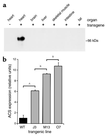
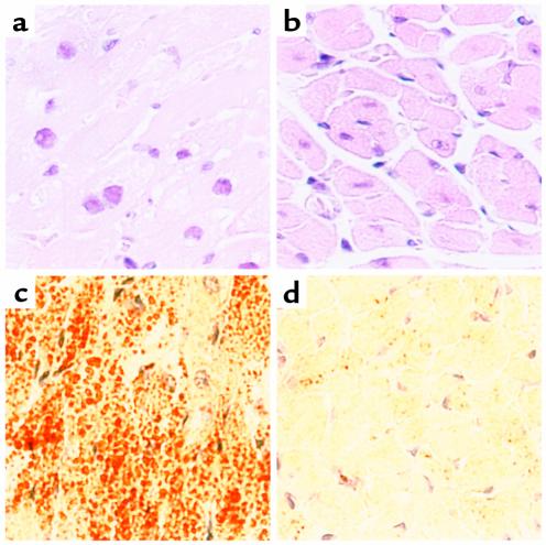
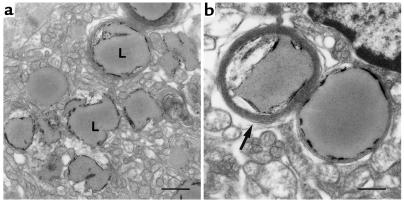
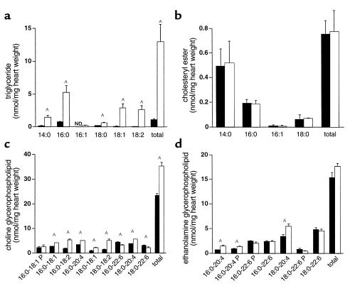
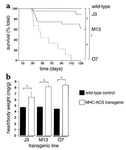
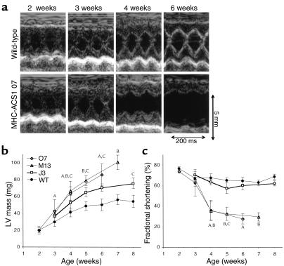
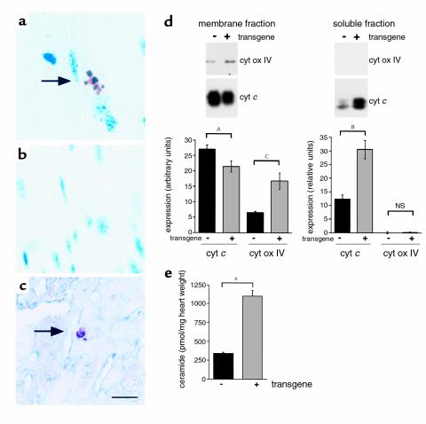
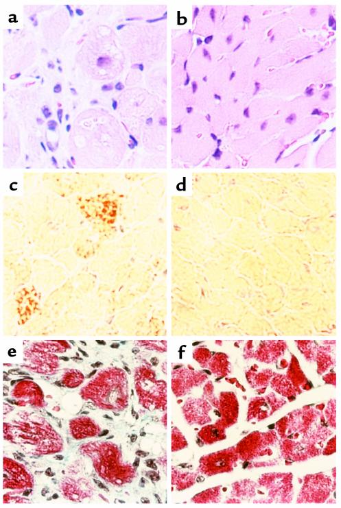
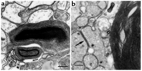
Inherited and acquired cardiomyopathies are associated with marked intracellular lipid accumulation in the heart. To test the hypothesis that mismatch between myocardial fatty acid uptake and utilization leads to the accumulation of cardiotoxic lipid species, and to establish a mouse model of metabolic cardiomyopathy, we generated transgenic mouse lines that overexpress long-chain acyl-CoA synthetase in the heart (MHC-ACS). This protein plays an important role in vectorial fatty acid transport across the plasma membrane. MHC-ACS mice demonstrate cardiac-restricted expression of the transgene and marked cardiac myocyte triglyceride accumulation. Lipid accumulation is associated with initial cardiac hypertrophy, followed by the development of left-ventricular dysfunction and premature death. Terminal deoxynucleotidyl transferase-mediated dUTP nick-end labeling staining and cytochrome c release in transgenic hearts suggest that cardiac myocyte death occurs, in part, by lipid-induced programmed cell death. Taken together, our data demonstrate that fatty acid uptake/utilization mismatch in the heart leads to accumulation of lipid species toxic to cardiac myocytes. This novel mouse model will provide insight into the role of perturbations in myocardial lipid metabolism in the pathogenesis of inherited and acquired forms of heart failure.


Figures

Figure 1
Figure 1
Cardiac overexpression of murine ACS1 in MHC-ACS lines. (a) Membrane protein (20 μg) from various organs of an MHC-ACS O7 transgenic animal and nontransgenic littermate were separated by SDS-PAGE. Specific transgene expression was analyzed by Western blot, using a monoclonal anti-MYC Ab. (b) Membrane protein (20 μg) from hearts of 18-day-old MHC-ACS transgenic mice were analyzed by Western blot using rabbit polyclonal antisera directed against native murine ACS1 sequences. ACS1-specific signal was quantified and relative units of ACS1 expression is shown for wild-type (WT) and three independent transgenic lines (J3, M13, and O7). Data are reported as the mean ± SE. Differences among groups were compared by one-way ANOVA in conjunction with the post hoc Scheffé test (AP < 0.0001; BP < 0.01).
Figure 2
Figure 2
Light-microscopic examination of cardiac tissue from 18-day-old MHC-ACS mice. Cardiac ventricular tissue was dissected from 18-day-old MHC-ACS O7 animals (a, c) and nontransgenic littermates (b, d). Tissue was fixed in formalin, embedded in paraffin, and sectioned for H&E staining (a, b). Tissue was flash-frozen and sectioned for oil red O staining (c, d). ×400.
Figure 3
Figure 3
EM of cardiac tissue from 18-day-old MHC-ACS mice. Ultra-thin sections of fixed ventricular tissue from 18-day-old MHC-ACS O7 animals were examined by transmission EM. Images are shown at ×7,500 (a; bar, 1 μm), and at ×15,000 (b; bar, 0.5 μm). Numerous lipid droplets (L) are observed in ventricular myocytes. Some droplets at this stage are surrounded by multiple concentric layers of membrane (arrow).
Figure 4
Figure 4
Cardiac lipid accumulation in 18-day-old MHC-ACS mice. Lipids were extracted from frozen ventricular tissues of wild-type and transgenic (O7) mice. Triacylglycerols (a), cholesteryl esters (b), choline glycerophospholipids (c), and ethanolamine glycerophospholipids (d), were quantified. Data from wild-type and transgenic mice are displayed with filled and open bars, respectively. Data for each measurement report the mean of a minimum of five animals ± SE. Statistical evaluation between groups was by Student’s t test (AP < 0.01). ND, not detected; P, plasmalogen species.
Figure 5
Figure 5
Premature death and cardiomegaly in MHC-ACS mice. (a) Wild-type (WT, n = 110) and transgenic (J3, n = 19; M13, n = 23; O7, n = 9) mice were followed for 130 days, and incidence of spontaneous death was recorded as a function of time. Differences among survival curves were compared using the log-rank test (AP < 0.05, BP < 0.01, CP < 0.0001). (b) At the time of spontaneous death or sacrifice, mice were weighed. Hearts were dissected, and atrial and vascular structures were removed before weighing. Heart-to-body weight ratios were determined in wild-type (solid bars) and transgenic (open bars for J3, M13, O7) mice older than 24 days at the time of death or sacrifice. For each transgenic mouse, an age- and sex-matched wild-type littermate was sacrificed and analyzed. Differences among groups were compared by two-way ANOVA (AP < 0.0001).
Figure 6
Figure 6
Cardiac hypertrophy and failure in MHC-ACS mice. (a) Two-dimensional guided M-mode echocardiographic images were obtained from wild-type (upper panel) and MHC-ACS1 O7 (lower panel) mice at 2, 3, 4, and 6 weeks of age. Images shown are from a representative pair of age- and sex-matched wild-type and transgenic animals. (b) Quantification of left-ventricular (LV) mass was performed on age- and sex-matched wild-type and transgenic (J3, M13, O7) mice based on serial echocardiograms at the indicated time points. A minimum of five different animals were examined at each time point. Data points are expressed as mean ± SD. A,B,CP < 0.05 for O7, M13, or J3, respectively, versus wild-type. (c) Quantification of fractional shortening over time as assessed by serial echocardiograms for age- and sex-matched wild-type and transgenic mice (J3, M13, O7). Data points are expressed as mean ± SD. A,B,CP < 0.05 for O7, M13, or J3, respectively, versus wild-type.
Figure 7
Figure 7
Evidence for lipoapoptosis in MHC-ACS hearts. (ac) Cardiac ventricular tissues from a 18-day-old O7 transgenic mice (a and c) and from a nontransgenic control littermate (b) were fixed in formalin, embedded in paraffin, and sectioned. Tissues were stained for DNA fragmentation by a TUNEL protocol that stains apoptotic nuclei brown and allows visualization of myocyte striations. Double staining for α-sarcomeric actin was used to identify cardiomyocytes in c (myocyte cytoplasm blue). (d) Cardiac ventricular tissues from 21-day-old O7 transgenic mice and nontransgenic control littermates were flash-frozen, homogenized, and separated by differential density centrifugation to yield a membrane fraction (mitochondria) and soluble fraction (cytosol). Membrane protein (20 μg) and soluble protein (80 μg) were separated by SDS-PAGE and analyzed by Western blotting using an anti–cytochrome c (cyt c) Ab and an anti–cytochrome oxidase subunit IV (cyt ox IV) Ab. Bands were quantified using Molecular Analyst software, and relative units of expression are shown for wild-type (–) and transgenic (+) tissues. Data points are expressed as mean (minimum of four independent samples) ± SE. Nonpaired t test was used to compare groups (AP = 0.06, BP = 0.01, CP < 0.01). (e) Ceramide was measured in heart tissue from 18-day-old transgenic and wild-type animals using the diacylglycerol kinase assay and normalized for tissue weight. Data points are expressed as mean (minimum of five independent samples) ± SE. Statistical evaluation between groups was by Student’s t test (AP < 0.0005).
Figure 8
Figure 8
Light-microscopic examination of cardiac tissue from end-stage MHC-ACS O7 mice. Cardiac ventricular tissue was dissected from 28-day-old MHC-ACS O7 animals (a, c, e) and nontransgenic littermates (b, d, f). Tissue was fixed in formalin, embedded in paraffin, and sectioned for H&E staining (a, b) or Masson’s trichrome (e, f). Tissue was flash-frozen and sectioned for oil red O staining (c, d). ×400.
Figure 9
Figure 9
EM of cardiac tissue from end-stage MHC-ACS O7 mice. Ventricular tissue from a 28-day-old transgenic mouse was fixed in formalin and thin sectioned for EM. ×15,000. Note complex structures composed of multiple layers of membrane in the cytoplasm of myocytes (a). In addition to these large multilamellar membrane aggregates, membranes often accumulated within the cytoplasm between organelles such as mitochondria (b) (arrow). Bar, 0.5 μm.

References

    1. Neely JR, Rovetto MJ, Oram JF. Myocardial utilization of carbohydrate and lipids. Prog Cardiovasc Dis. 1972;15:289–329. - PubMed
    1. van der Vusse GJ, Glatz JF, Stam HC, Reneman RS. Fatty acid homeostasis in the normoxic and ischemic heart. Physiol Rev. 1992;72:881–940. - PubMed
    1. Richieri GV, Kleinfeld AM. Unbound free fatty acid levels in human serum. J Lipid Res. 1995;36:229–240. - PubMed
    1. Stremmel W. Fatty acid uptake by isolated rat heart myocytes represents a carrier-mediated transport process. J Clin Invest. 1988;81:844–852. - PMC - PubMed
    1. Sorrentino D, Robinson RB, Kiang C-L, Berk PD. At physiologic albumin/oleate concentrations oleate uptake by isolated hepatocytes, cardiac myocytes, and adipocytes is a saturable function of the unbound oleate concentration. J Clin Invest. 1989;84:1325–1333. - PMC - PubMed

Publication types

Substances