. 2001 Apr;107(7):871-80.
doi: 10.1172/JCI11708.

The regulatory role of DR4 in a spontaneous diabetes DQ8 transgenic model

Affiliations

The regulatory role of DR4 in a spontaneous diabetes DQ8 transgenic model

L Wen et al. J Clin Invest. 2001 Apr.

Abstract

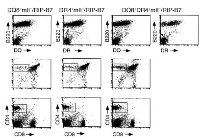
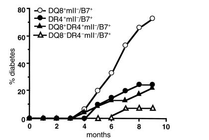
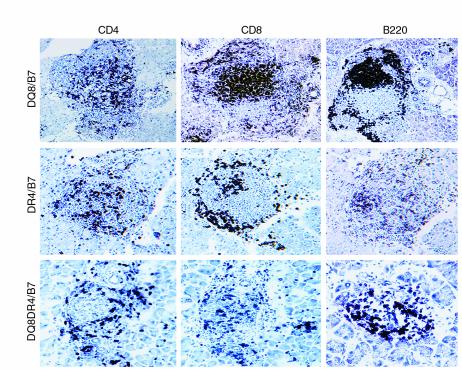
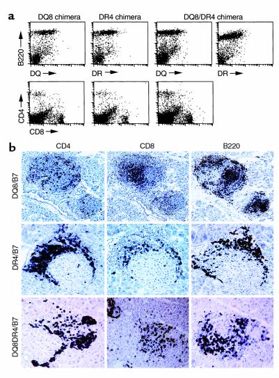
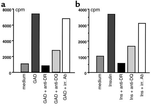
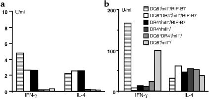
MHC class II molecules are critical determinants of genetic susceptibility to human type 1 diabetes. In patients, the most common haplotype contains the DRA1*0101-DRB1*0401 (DR4) and DQA1*0301-DQB1*0302 (DQ8) loci. To assess directly the relative roles of HLA-DQ8 and DR4 for diabetes development in vivo, we generated C57BL/6 transgenic mice that lack endogenous mouse MHC class II molecules but express HLA-DQ8 and/or DR4. Neither HLA-DQ nor HLA-DR transgenic mice developed insulitis or spontaneous diabetes. However, when they were crossed to transgenic mice (C57BL/6) expressing the B7.1 costimulatory molecules on pancreatic beta cells that do not normally develop diabetes, T cells from these double transgenic mice were no longer tolerant to islet autoantigens. The majority of DQ8/RIP-B7 mice developed spontaneous diabetes, whereas only 25% of DR4/RIP-B7 mice did so. Interestingly, when DQ8 and DR4 were coexpressed (DQ8DR4/RIP-B7), only 23% of these mice developed diabetes, an incidence indistinguishable from the DR4/RIP-B7 mice. T cells from both DR4/RIP-B7 and DQ8DR4/RIP-B7 mice, unlike those from DQ8/RIP-B7 mice, exhibited a Th2-like phenotype. Thus, the expression of DR4 appeared to downregulate DQ8-restricted autoreactive T cells in DQ8DR4/RIP-B7 mice. Our data suggest that although both DQ8 and DR4 can promote spontaneous diabetes in mice with a non-autoimmune-prone genetic background, the diabetogenic effect of the DQ8 allele is much greater, whereas DR4 expression downregulates the diabetogenic effect of DQ8, perhaps by enhancing Th2-like immune responses.


Figures

Figure 1
Figure 1
Expression of HLA transgenes and the selection of CD4+ T cells. In the upper panel, lymphocytes isolated from peripheral blood in three types of mice (as indicated) were stained with the B-cell marker B220 (phycoerythrin; PE) in combination with either anti–HLA-DQ (FITC) or DR (FITC). In the middle panel, thymocytes were isolated from mice at 5–7 weeks of age and stained with anti-CD4 (PE; y axis) and anti-CD8 (FITC; x axis). Gated CD4 single-positive thymocytes were 5.2%, 5%, and 9.8% of total thymocytes analyzed in DQ8+/mII/RIP-B7, DR4+/mII/RIP-B7, and DQ8+DR4+/mII/RIP-B7 mice, respectively. In the bottom panel, splenocytes (after removing erythrocytes) were stained with anti-CD4 (PE) and anti-CD8 (FITC). Gated CD4 single-positive splenocytes were 11.4%, 11.8%, and 19.5% of total splenocytes analyzed in DQ8+/mII/RIP-B7, DR4+/mII/RIP-B7, and DQ8+DR4+/mII/RIP-B7 mice, respectively.
Figure 2
Figure 2
Spontaneous diabetes development in HLA/RIP-B7 transgenic mice. Four groups of mice were used in the study as indicated. Number of mice per group was: n = 15 (7 females and 8 males) for DQ8+/mII/RIP-B7; n = 20 (11 females and 9 males) for DR4+/mII/RIP-B7; n = 22 (12 females and 10 males) for DQ8+DR4+/mII/RIP-B7; n = 10 (4 females and 6 males) for mII/RIP-B7 mice. Mice were housed in specific pathogen-free (SPF) conditions. Diabetes was determined by monitoring of urinary glucose and confirmed by blood glucose (>250 mg/dl).
Figure 3
Figure 3
Immunohistochemistry staining of pancreatic sections of diabetic mice, with the diabetic DQ8+/mII/RIP-B7 mouse in the upper panel, diabetic DR4+/mII/RIP-B7 mouse in the middle panel, and diabetic DQ8+DR4+/mII/RIP-B7 mouse in the lower panel. Islet infiltrates were stained for CD4+ T cells (left), CD8+ T cells (middle), and B220+ B cells (right).
Figure 4
Figure 4
(a) Expression of HLA transgenes and the selection of CD4+ T cells in BM chimeras. In the top panel, lymphocytes isolated from the spleens of three types of chimeras (as indicated) were stained with the B-cell marker B220 (PE) in combination with either anti–HLA-DQ (FITC) or DR (FITC). In the lower panel, the same splenocytes were stained with anti-CD4 (PE) and anti-CD8 (FITC). (b) Immunohistochemistry staining of pancreatic sections of BM chimeras. In the upper panel are DQ8+/mII/RIP-B7 BM chimera. DR4+/mII/RIP-B7 BM chimera are in the middle panel. The lower panel shows DQ8+DR4+/mII/RIP-B7 BM chimera. Islet infiltrates were stained for CD4+ T cells (left), CD8+ T cells (middle), and B220+ B cells (right).
Figure 5
Figure 5
In vitro proliferation of T cells derived from diabetic spleens to islet β-cell autoantigens GAD and insulin (Ins). (a) Purified T cells (2 × 10⁵/well) were cultured in medium (Click’s medium containing 5% heat-inactivated FCS) alone or with Ag’s (as indicated) in the presence of irradiated (irr.) splenocytes as Ag-presenting cells. (b) HLA restriction of the proliferative response was also tested in the assays by addition of supernatants of mAb’s to DR (HB55) and DQ (HB144). The irrelevant control Ab used in the assays was anti-IAb (Y3JP). The cultures were pulsed with 0.5 μCi 3H after 72 hours.
Figure 6
Figure 6
Cytokine production by CD4+ T cells after GAD (a) and anti-CD3 (b) stimulation. Purified CD4+ cells (2 × 10⁶/ml) from spleens of nondiabetic mice, as indicated, were cultured with GAD protein or anti-CD3 (2C11 supernatant) in the presence of irradiated splenocytes for 72 and 48 hours, respectively. The culture supernatants were then tested for the production of IFN-γ and IL-4 using ELISA.
Figure 7
Figure 7
The mRNA expression of chemokines CCR5 and CCR2. Expression of chemokines CCR5 and CCR2 mRNA was assessed using RT-PCR (see Methods), as shown in the ethidium-bromide stained gel. CD3ε was used as the internal control for the normalization of RNA levels of tested samples, as indicated. Lane 1: 100-bp ladder; lanes 2–5: CCR5 or CCR2, as shown. RT-PCR was performed on oligo-dT–synthesized cDNA, which was derived from total RNA of anti-CD3–stimulated T cells from spleens of nondiabetic mice as indicated.
Figure 8
Figure 8
Expression of Ig isotypes in DQ8+/mII/RIP-B7, DR4+/mII/RIP-B7, and DQ8+DR4+/mII/RIP-B7 mice. Serum levels of IgG1 and IgA were measured using ELISA (see Methods) in the three groups of mice as indicated. The number of mice in each group was 15, 20, and 22 for DQ8+/mII/RIP-B7, DR4+/mII/RIP-B7, and DQ8+DR4+/mII/RIP-B7 mice, respectively. The results are presented as mean ± SD. Student’s t test was used for the statistical analysis.

Comment in

  • DR, DQ, and you: MHC alleles and autoimmunity.
    Sønderstrup G, McDevitt HO. Sønderstrup G, et al. J Clin Invest. 2001 Apr;107(7):795-6. doi: 10.1172/JCI12634. J Clin Invest. 2001. PMID: 11285296 Free PMC article. Review. No abstract available.

Similar articles

Cited by

References

    1. Davies JL, et al. A genome-wide search for human type 1 diabetes susceptibility genes. Nature. 1994;371:130–136. - PubMed
    1. Tisch R, McDevitt H. Insulin-dependent diabetes mellitus. Cell. 1996;85:291–297. - PubMed
    1. Todd JA, Farrall M. Panning for gold: genome-wide scanning for linkage in type 1 diabetes. Hum Mol Genet. 1996;5:1443–1448. - PubMed
    1. Bendelac A, Carnaud C, Boitard C, Bach JF. Syngeneic transfer of autoimmune diabetes from diabetic NOD mice to healthy neonates. Requirement for both L3T4+ and Lyt-2+ T cells. J Exp Med. 1987;166:823–832. - PMC - PubMed
    1. Miller BJ, Appel MC, O’Neil JJ, Wicker LS. Both the Lyt-2+ and L3T4+ T cell subsets are required for the transfer of diabetes in nonobese diabetic mice. J Immunol. 1988;140:52–58. - PubMed

Publication types

MeSH terms