

West Nile virus recombinant DNA vaccine protects mouse and horse from virus challenge and expresses in vitro a noninfectious recombinant antigen that can be used in enzyme-linked immunosorbent assays
- PMID: 11287553
- PMCID: PMC114149
- DOI: 10.1128/JVI.75.9.4040-4047.2001
West Nile virus recombinant DNA vaccine protects mouse and horse from virus challenge and expresses in vitro a noninfectious recombinant antigen that can be used in enzyme-linked immunosorbent assays
Abstract
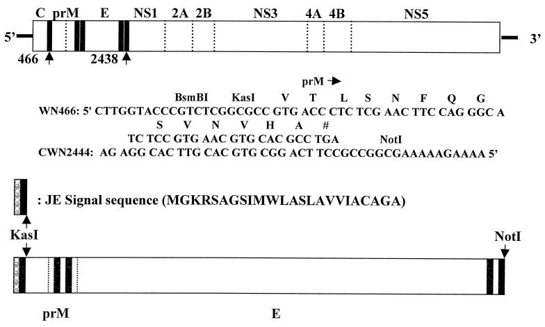
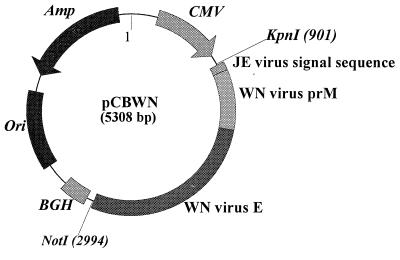
Introduction of West Nile (WN) virus into the United States in 1999 created major human and animal health concerns. Currently, no human or veterinary vaccine is available to prevent WN viral infection, and mosquito control is the only practical strategy to combat the spread of disease. Starting with a previously designed eukaryotic expression vector, we constructed a recombinant plasmid (pCBWN) that expressed the WN virus prM and E proteins. A single intramuscular injection of pCBWN DNA induced protective immunity, preventing WN virus infection in mice and horses. Recombinant plasmid-transformed COS-1 cells expressed and secreted high levels of WN virus prM and E proteins into the culture medium. The medium was treated with polyethylene glycol to concentrate proteins. The resultant, containing high-titered recombinant WN virus antigen, proved to be an excellent alternative to the more traditional suckling-mouse brain WN virus antigen used in the immunoglobulin M (IgM) antibody-capture and indirect IgG enzyme-linked immunosorbent assays. This recombinant antigen has great potential to become the antigen of choice and will facilitate the standardization of reagents and implementation of WN virus surveillance in the United States and elsewhere.
Figures
References
-
- Aberle J H, Aberle S W, Allison S L, Stiasny K, Ecker M, Mandl C W, Berger R, Heinz F X. A DNA immunization model study with constructs expressing the tick- borne encephalitis virus envelope protein E in different physical forms. J Immunol. 1999;163:6756–6761. - PubMed
-
- Anderson J F, Andreadis T G, Vossbrinck C R, Tirrell S, Wakem E M, French R A, Garmendia A E, Van Kruiningen H J. Isolation of West Nile virus from mosquitoes, crows, and a Cooper's hawk in Connecticut. Science. 1999;286:2331–2333. - PubMed
-
- Anonymous. Update: surveillance for West Nile virus in overwintering mosquitoes —New York, 2000. Morb Mortal Wkly Rep. 2000;49:178–179. - PubMed
MeSH terms
Substances
LinkOut - more resources
Full Text Sources
Other Literature Sources
Medical
