. 2001 May;75(9):4184-94.
doi: 10.1128/JVI.75.9.4184-4194.2001.

Polyadenylation in rice tungro bacilliform virus: cis-acting signals and regulation

Affiliations

Polyadenylation in rice tungro bacilliform virus: cis-acting signals and regulation

H M Rothnie et al. J Virol. 2001 May.

Abstract

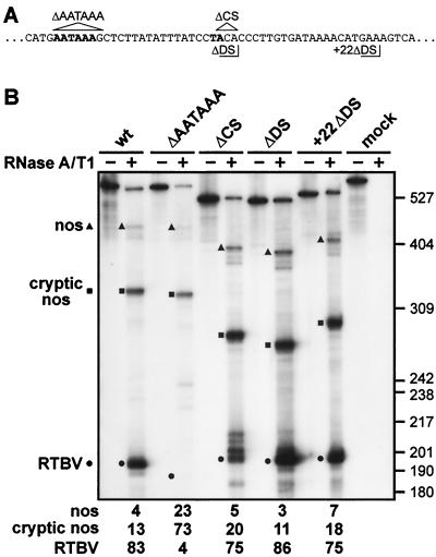
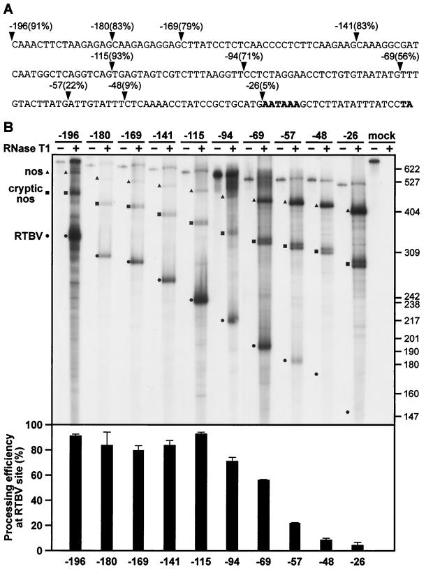
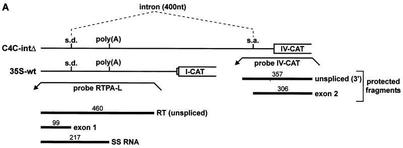
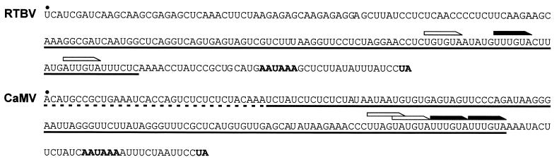
The polyadenylation signal of rice tungro bacilliform virus (RTBV) was characterized by mutational and deletion analysis. The cis-acting signals required to direct polyadenylation conformed to what is known for plant poly(A) signals in general and were very similar to those of the related cauliflower mosaic virus. Processing was directed by a canonical AAUAAA poly(A) signal, an upstream UG-rich region considerably enhanced processing efficiency, and sequences downstream of the cleavage site were not required. When present at the end of a transcription unit, the cis-acting signals for 3'-end processing were highly efficient in both monocot (rice) and dicot (Nicotiana plumbaginifolia) protoplasts. In a promoter-proximal position, as in the viral genome, the signal was also efficiently processed in rice protoplasts, giving rise to an abundant "short-stop" (SS-) RNA. The proportion of SS-RNA was considerably lower in N. plumbaginifolia protoplasts. In infected plants, SS-RNA was hardly detectable, suggesting either that SS-RNA is unstable in infected plants or that read-through of the promoter-proximal poly(A) site is very efficient. SS-RNA is readily detectable in transgenic rice plants (A. Klöti, C. Henrich, S. Bieri, X. He, G. Chen, P. K. Burkhardt, J. Wünn, P. Lucca, T. Hohn, I. Potrylus, and J. Fütterer, 1999. Plant Mol. Biol. 40:249-266), thus the absence of SS-RNA in infected plants can be attributed to poly(A) site bypass in the viral context to ensure production of the full-length pregenomic viral RNA. RTBV poly(A) site suppression thus depends both on context and the expression system; our results suggest that the circular viral minichromosome directs assembly of a transcription-processing complex with specific properties to effect read-through of the promoter-proximal poly(A) signal.


Figures

FIG. 1
FIG. 1
(A) Genomic map of RTBV and experimental strategy. The lower part of the figure shows the genome map of RTBV. Viral DNA is represented by a double line, with the box marked R′ indicating the region of the genome that is transcribed twice in the terminally redundant transcript. The thick arrows outside the DNA represent the major viral ORFs (I through IV). Viral transcripts are shown as thin arrows inside the DNA, with the 6.3-kb intron in the spliced transcript encoding ORF IV indicated (dashed line). The basic expression plasmid is represented schematically in the upper part of the panel. The 35S promoter, CAT reporter gene, and RTBV/nos sequences are represented as open boxes. The RTBV, cryptic nos, and nos cleavage sites are indicated with a solid arrow, an open arrowhead, and an open arrow, respectively. The position of the antisense probe transcript for RNase protection analysis is indicated. Homologous probes were used for each construct and were transcribed from linearized plasmid using the T7 promoter present downstream of the nos sequence in the vector. The RTBV sequences inserted in RTPA-L and RTPA-S are indicated with the numbers referring to the transcription start site. Processing efficiencies are given in percent and were roughly the same in N. plumbaginifolia and rice protoplasts, with values from cryptic nos and nos sites being combined as “read through.” Values given are the average of at least three independent transfections. (B) Representative RNase protection assays of RNAs expressed from constructs RTPA-L and RTPA-S. Fragments corresponding to processing at the RTBV, cryptic nos, and nos sites are indicated with a solid arrow, an open arrowhead, and an open arrow, respectively. Signal intensities were always weaker from rice protoplast RNA (RTPA-L not shown). The positions of labeled DNA size markers (pBR322/HpaII) are indicated. The expected sizes of protected fragments at the RTBV, cryptic nos, and nos sites are 515, 818, and 932 nt, or 211, 334, and 448 nt with the RTPA-L and RTBV-S probes, respectively. Signals at the size of the full-length probe in this and other figures are discussed in the text.
FIG. 2
FIG. 2
AATAAA is required for 3′-end processing; the cleavage site and specific downstream sequences are not. (A) Sequences surrounding the AATAAA and cleavage sites (both in bold type): nucleotides deleted in ΔAATAAA and ΔCS are indicated by triangles; the end points of RTBV sequences in ΔDS and +22ΔDS are delimited by right-angled lines. (B) RNase protection assay to examine processing events at the RTBV poly(A) site in a wild-type construct (RTPA-2), and constructs carrying deletions of either AATAAA (ΔAATAAA), the cleavage site (ΔCS), or sequences directly downstream of the cleavage site (ΔDS) or from 22 nt downstream of the cleavage site (+22ΔDS) as shown in panel A. Protected fragments corresponding to transcripts processed at the RTBV, cryptic nos, and nos sites are indicated, with the positions of size markers (pBR322/HpaII) shown on the right. Processing efficiencies (in percent) are given underneath. The gel shown is from analysis of RNA from N. plumbaginifolia protoplasts.
FIG. 3
FIG. 3
Sequences upstream of AATAAA enhance processing efficiency at the RTBV site. (A) The RTBV sequence in RTPA-S up to the site of poly(A) addition is shown. Arrowheads mark the endpoints of the ExoIII deletion series, with the number of nucleotides remaining upstream of the cleavage site and the corresponding processing efficiency indicated. (B) Representative RNase T1 protection assay of the deletion series shown in panel A. Protected fragments corresponding to transcripts processed at the RTBV, cryptic nos, and nos sites are indicated, with the positions of size markers (pBR322/HpaII) shown on the right. Processing efficiencies at the RTBV site are shown in the chart underneath (error bars represent standard deviations). The gel shown is from analysis of RNA from N. plumbaginifolia protoplasts.
FIG. 4
FIG. 4
Processing at the RTBV poly(A) site in a promoter-proximal position gives rise to SS-RNA in transfected protoplasts. (A) Constructs consisting of the RTBV (to −681) or CaMV 35S (to −343) upstream promoter sequences, the RTBV leader sequence (either complete [wt] or deleted between +8 and +83 [Δ]) and the CAT reporter gene fused to RTBV ORF I are shown. All constructs are terminated by the CaMV polyadenylation signal. The location of the homologous region of the antisense probe used for RNase A/T1 mapping, and the extent of protected fragments corresponding to SS and RT transcripts, are indicated. (B and C) RNase protection analysis. Total RNA isolated from transfected rice (B) or N. plumbaginifolia (C) protoplasts was subjected to RNase A/T1 protection analysis with an antisense probe transcribed from RTPA-L. Protected fragments corresponding to RTBV SS and RT transcripts for the four constructs used are indicated, with the percentage of SS shown below the gels. ∗, Values with the RTBV-Δ construct in N. plumbaginifolia were too low to quantify reliably. Other values given are the average from two independent experiments (variation was within 10% of the mean). i.c., internal control; pDES7 (see Materials and Methods).
FIG. 5
FIG. 5
SS-RNA is not detected in RTBV-infected rice plants. (A) The RTBV pgRNA is represented twice, by solid and dashed curved lines, with the SS-RNA as a straight line colinear with the 3′ end of the pgRNA. The antisense probes transcribed from RTPA-L or RTPA-2 are represented above the upper and lower diagrams, respectively, and thick lines indicate the position and expected sizes (in nucleotides) of protected RNA fragments corresponding to the 5′ and 3′ ends of the pgRNA and the SS-RNA. The diagram is not to scale, and the nos sequences on the probes are not indicated. (B) RNase protection analysis of total RNA (5-μg aliquots) from RTBV-infected rice plants using probes covering the terminal redundancy (RTPA-L), the 3′ end of the terminal redundancy (RTPA-2), or a 135-nt fragment of the RTBV pgRNA spanning the end of ORF III and the beginning of ORF IV (IV-CAT [10] protects a 135-nt internal fragment on the pgRNA). Fragments obtained with probe RTPA-L corresponding to the 5′ (RT) and 3′ ends of the RTBV pgRNA are indicated on the left, as well as the expected position of SS-RNA. A longer fragment (RT2) is also indicated (see text for possible explanations). Fragments protected by the RTPA-2 and IV-CAT probes are labeled on the right. Note that the RTPA-2 probe does not distinguish between SS-RNA and the 3′ end of pgRNA.
FIG. 6
FIG. 6
SS-RNA is unaffected by splicing of the viral intron. (A) Constructs C4C-intΔ and 35S-wt are depicted schematically, with the positions of the splice donor (s.d.), splice acceptor (s.a.), and poly(A) site indicated. Splicing of the 400-nt intron is indicated by dashed lines. Probes RTPA-L and IV-CAT are shown as leftward pointing arrows. The position and size (in nucleotides) of protected fragments are shown underneath. (B) Representative RNase protection analyses of the constructs shown in panel A, expressed in either rice (left panel) or N. plumbaginifolia (right panel) protoplasts. Probes RTPA-L and IV-CAT were used together in each sample. Protected fragments are labeled and named as in panel A. In rice protoplasts, some splicing events occurred on the 35S-wt transcript, presumably using cryptic acceptor sites within the CAT ORF or in the vector sequences. (C) The amount of SS-RNA in percent (calculated as SS/[SS+RT+exon 1]).
FIG. 6
FIG. 6
SS-RNA is unaffected by splicing of the viral intron. (A) Constructs C4C-intΔ and 35S-wt are depicted schematically, with the positions of the splice donor (s.d.), splice acceptor (s.a.), and poly(A) site indicated. Splicing of the 400-nt intron is indicated by dashed lines. Probes RTPA-L and IV-CAT are shown as leftward pointing arrows. The position and size (in nucleotides) of protected fragments are shown underneath. (B) Representative RNase protection analyses of the constructs shown in panel A, expressed in either rice (left panel) or N. plumbaginifolia (right panel) protoplasts. Probes RTPA-L and IV-CAT were used together in each sample. Protected fragments are labeled and named as in panel A. In rice protoplasts, some splicing events occurred on the 35S-wt transcript, presumably using cryptic acceptor sites within the CAT ORF or in the vector sequences. (C) The amount of SS-RNA in percent (calculated as SS/[SS+RT+exon 1]).
FIG. 7
FIG. 7
Comparison of the poly(A) signals of RTBV and CaMV. The sequences of the terminal redundancies of RTBV and CaMV are shown. The region is delimited by the transcription start site (black dot) and the poly(A) site (bold type UA). NUEs are set in bold type, and FUEs are underlined, with a dashed underline indicating sequences with only a slight effect on processing efficiency. Perfect and one-base mismatches of UUUGUA are indicated with black and white arrows, respectively. (Adapted from reference .)

Similar articles

Cited by

References

    1. Ashe M P, Furger A, Proudfoot N J. Stem-loop 1 of the U1 snRNP plays a critical role in the suppression of HIV-1 polyadenylation. RNA. 2000;6:170–177. - PMC - PubMed
    1. Ashe M P, Pearson L H, Proudfoot N J. The HIV-1 5′ poly(A) site is inactivated by U1 snRNP interaction with the downstream major splice donor site. EMBO J. 1997;16:5752–5763. - PMC - PubMed
    1. Bao Y, Hull R. Mapping the 5′-terminus of rice tungro bacilliform viral genomic RNA. Virology. 1993;197:445–448. - PubMed
    1. Bentley D. Coupling RNA polymerase II transcription with pre-mRNA processing. Curr Opin Cell Biol. 1999;11:347–351. - PubMed
    1. Brackenridge S, Proudfoot N J. Recruitment of a basal polyadenylation factor by the upstream sequence element of the human lamin B2 polyadenylation signal. Mol Cell Biol. 2000;20:2660–2669. - PMC - PubMed

LinkOut - more resources