. 2001 May;75(9):4247-57.
doi: 10.1128/JVI.75.9.4247-4257.2001.

Hepatitis B virus HBx protein activation of cyclin A-cyclin-dependent kinase 2 complexes and G1 transit via a Src kinase pathway

Affiliations

Hepatitis B virus HBx protein activation of cyclin A-cyclin-dependent kinase 2 complexes and G1 transit via a Src kinase pathway

M Bouchard et al. J Virol. 2001 May.

Abstract

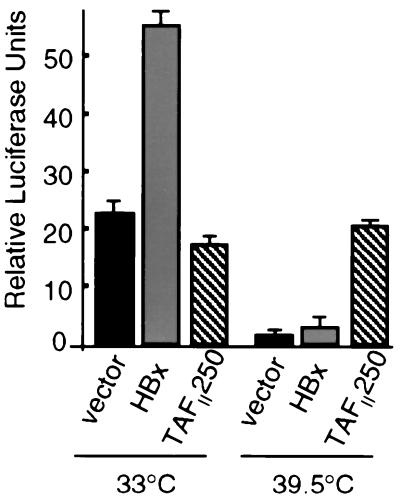
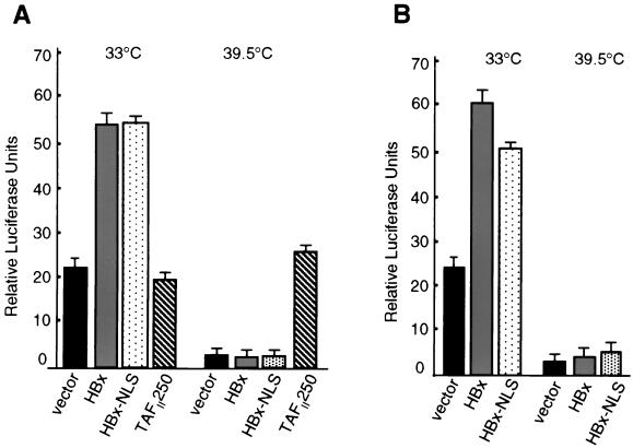
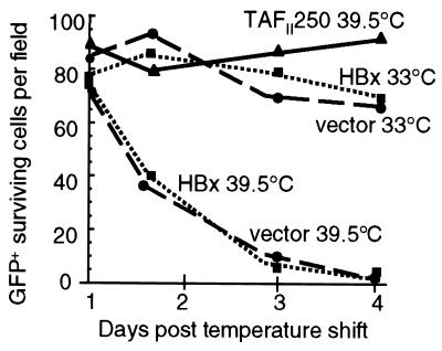
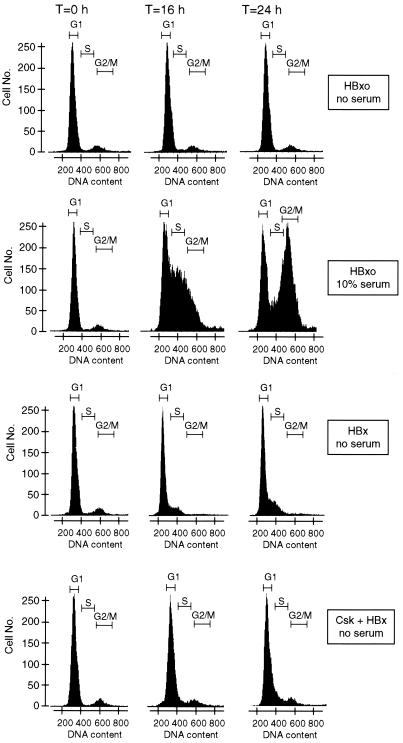
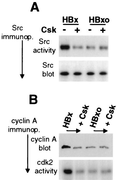
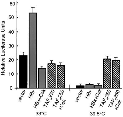
Numerous studies have demonstrated that the hepatitis B virus HBx protein stimulates signal transduction pathways and may bind to certain transcription factors, particularly the cyclic AMP response element binding protein, CREB. HBx has also been shown to promote early cell cycle progression, possibly by functionally replacing the TATA-binding protein-associated factor 250 (TAF(II)250), a transcriptional coactivator, and/or by stimulating cytoplasmic signal transduction pathways. To understand the basis for early cell cycle progression mediated by HBx, we characterized the molecular mechanism by which HBx promotes deregulation of the G0 and G1 cell cycle checkpoints in growth-arrested cells. We demonstrate that TAF(II)250 is absolutely required for HBx activation of the cyclin A promoter and for promotion of early cell cycle transit from G0 through G1. Thus, HBx does not functionally replace TAF(II)250 for transcriptional activity or for cell cycle progression, in contrast to a previous report. Instead, HBx is shown to activate the cyclin A promoter, induce cyclin A-cyclin-dependent kinase 2 complexes, and promote cycling of growth-arrested cells into G1 through a pathway involving activation of Src tyrosine kinases. HBx stimulation of Src kinases and cyclin gene expression was found to force growth-arrested cells to transit through G1 but to stall at the junction with S phase, which may be important for viral replication.


Figures

FIG. 1
FIG. 1
HBx stimulation of cyclin A promoter requires TAFII250 function. ts13 cells grown at 33°C were transiently transfected with control vector, HBx expression vector, or wild-type TAFII250 expression vector plus a cyclin A promoter-luciferase reporter construct. Cells were maintained at the permissive temperature (33°C) or shifted 5 h later to the nonpermissive temperature (39.5°C) for TAFII250 activity as described previously (24). At 24 h posttransfection, transcriptional activity was measured by relative luciferase light units and normalized for transfection efficiency and protein concentration. Data represent the means of at least three independent experiments, with calculated standard errors shown.
FIG. 2
FIG. 2
HBx remains predominantly cytoplasmic in ts13 cells regardless of temperature. ts13 cells were grown on coverslips at 33°C, transiently transfected with a plasmid expressing HBxFlag, which is an HBx protein containing a C-terminal Flag epitope that behaves identically to wild-type HBx, or HBxFlag containing an NLS (HBxNLS) or a control plasmid expressing a mutant NLS (HBxSLN) (23). Cells were maintained at 33°C or shifted to 39.5°C for 24 h, fixed and permeabilized on coverslips, and reacted with M2 anti-Flag antibodies followed by FITC-conjugated secondary antibody to visualize HBxFlag. Immunofluorescence photomicrographs (magnification, ×400) are shown for cells representative of each field. Control cells with vector alone did not demonstrate antibody staining (, ; data not shown).
FIG. 3
FIG. 3
Nuclear HBx does not complement for loss of TAFII250 in cyclin A or CRE-dependent promoter activity. (A) ts13 cells were transiently transfected with a cyclin A promoter-luciferase reporter construct and a vector expressing wild-type HBx, an HBx engineered to concentrate largely but not completely in the nucleus by inclusion of the SV40 T-Ag NLS (HBxNLS) (23), or wild-type TAFII250. (B) Cells were transfected as described above but with a 4× multimerized CRE-basal promoter-luciferase reporter construct. Cells were maintained at 33°C or shifted to the nonpermissive temperature for TAFII250 (39.5°C) for 24 h, and transcriptional activity was determined as described in the legend for Fig. 1. Results represent the means of at least three independent experiments, with calculated standard errors shown.
FIG. 4
FIG. 4
Effect of HBx on ts13 cell viability and growth in the absence of TAFII250 activity. ts13 cells at 33°C were transiently transfected with vector alone or with an HBx or wild-type TAFII250 expression vector. Duplicate plates of cells containing either vector alone or HBx were maintained at the permissive (33°C) temperature for endogenous TAFII250 activity. Cell viability and growth were determined at days 1 to 4 after the shift to the nonpermissive temperature based on cotransfected cells expressing a GFP marker, as described in Materials and Methods. Analysis of cell viability commenced at 24 h following the shift of samples to the nonpermissive temperature. Data represent a typical experiment that did not differ from three other studies by more than 10% and are displayed as the mean numbers of surviving cells per field obtained by the average of 10 fields per time point. An equal number of cells per field was plated at the start of the experiment.
FIG. 5
FIG. 5
Effect of HBx on the cell cycle. Change cells were accumulated largely in the G0-G1 phase by 30 h of maintenance in serum-free medium and then transduced with replication-defective Ad vectors with the wild-type HBx or mutant HBxo gene substituted for region E1. HBxo is a control that cannot synthesize HBx protein. Control cells containing HBxo were supplemented with 10% serum at the time of Ad transduction. Flow cytometry was performed using propidium iodide staining of nuclei. Histograms of nuclei are shown, obtained from cells at time zero (immediately following Ad transduction) and at 16 and 24 h. The DNA content of the nuclei was determined within 2 h of cell lysis by flow cytometry using the MODFIT program. Data are presented from a single experiment which did not vary by more than 10% in three trials.
FIG. 6
FIG. 6
HBx stimulates endogenous cyclin A promoter and cyclin A-cdk2 complexes through a Src kinase pathway. Quiescent serum-starved Chang cells were transiently transfected at ∼70% efficiency (data not shown) with vectors expressing HBx or HBxo, with and without cotransfection of a plasmid expressing Csk, a negative regulator of Src kinases. At 24 h posttransfection, cell lysates were prepared. (A) pp60 c-Src was immunoprecipitated from equal amounts of cell lysates using a specific monoclonal antibody, and equal fractions were resolved by SDS-PAGE (12% gel) and immunoblotted with the same antibody (Src blot) or assayed for in vitro phosphorylation activity with [γ-32P]ATP and the substrate enolase (Src activity). (B) Cyclin A was immunoprecipitated using a specific monoclonal antibody from equal amounts of lysate, and equal fractions of the immunoprecipitate were resolved by SDS-PAGE (15% gel) and immunoblotted for cyclin A protein (cyclin A blot) or assayed for associated cdk2 activity by in vitro phosphorylation of histone H1, a substrate of cdk2, using [γ-32P]ATP. Phosphorylated enolase and histone H1 were resolved by SDS-PAGE (12% gel), autoradiographed, and quantitated by digital densitometry. Results shown are typical of three independent experiments which did not vary by more than 20%.
FIG. 7
FIG. 7
HBx stimulation of cyclin A promoter through a Src kinase pathway. ts13 cells grown at the permissive temperature (33°C) or the restrictive temperature (39.5°C) were transiently transfected with the cyclin A promoter-luciferase reporter construct and control vector, HBx, or wild-type TAFII250, with or without a cotransfected Csk expression vector. Cells were then maintained at 33°C or shifted to 39.5°C 5 h after transcription. In some studies, cells were shifted to 39.5°C 24 h after transfection at 33°C, with identical results (data not shown). At 24 h posttransfection, transcriptional activity was measured by relative luciferase light units and normalized for transfection efficiency and protein concentration. Data represent the means of at least three independent experiments, with calculated standard errors shown.

References

    1. Albright S R, Tjian R. TAFs revisited: more data reveal new twists and confirm old ideas. Gene. 2000;242:1–13. - PubMed
    1. Andrisani O, Barnabas S. The transcriptional function of the hepatitis B virus X protein and its role in hepatocarcinogenesis. Int J Oncol. 1999;15:1–7. - PubMed
    1. Aufiero B, Schneider R J. The hepatitis B virus X-gene product trans-activates both RNA polymerase II and III promoters. EMBO J. 1990;9:497–504. - PMC - PubMed
    1. Barnabas S, Hai T, Andrisani O M. The hepatitis B virus X protein enhances the DNA binding potential and transcription efficacy of bZip transcription factors. J Biol Chem. 1997;272:20684–20690. - PubMed
    1. Barone M V, Courtneidge S A. Myc but not Fos rescue of PDGF signalling block caused by kinase-inactive Src. Nature. 1995;378:509–512. - PubMed

Publication types

MeSH terms

LinkOut - more resources