. 2001 May;75(9):4308-20.
doi: 10.1128/JVI.75.9.4308-4320.2001.

Regulation of human immunodeficiency virus type 1 infection, beta-chemokine production, and CCR5 expression in CD40L-stimulated macrophages: immune control of viral entry

Affiliations

Regulation of human immunodeficiency virus type 1 infection, beta-chemokine production, and CCR5 expression in CD40L-stimulated macrophages: immune control of viral entry

R L Cotter et al. J Virol. 2001 May.

Abstract

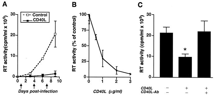
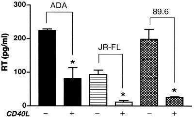
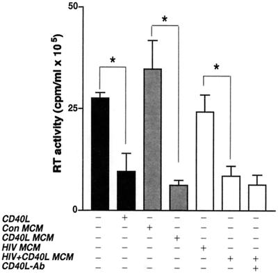
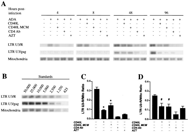
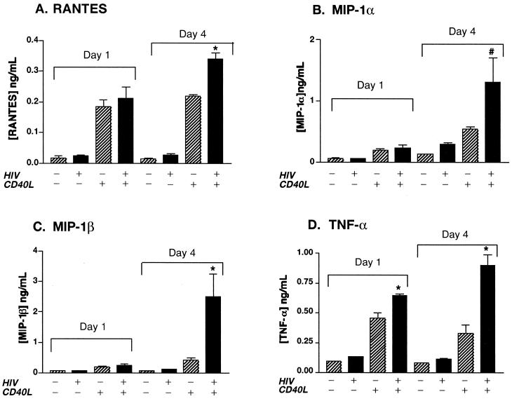
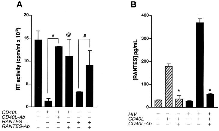
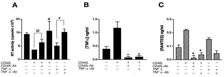
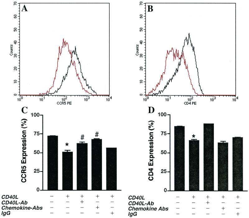
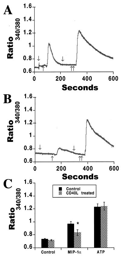
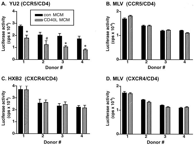
Mononuclear phagocytes (MP) and T lymphocytes play a pivotal role in the host immune response to human immunodeficiency virus type 1 (HIV-1) infection. Regulation of such immune responses can be mediated, in part, through the interaction of the T-lymphocyte-expressed molecule CD40 ligand (CD40L) with its receptor on MP, CD40. Upregulation of CD40L on CD4+ peripheral blood mononuclear cells during advanced HIV-1 disease has previously been reported. Based on this observation, we studied the influence of CD40L-CD40 interactions on MP effector function and viral regulation in vitro. We monitored productive viral infection, cytokine and beta-chemokine production, and beta-chemokine receptor expression in monocyte-derived macrophages (MDM) after treatment with soluble CD40L. Beginning 1 day after infection and continuing at 3-day intervals, treatment with CD40L inhibited productive HIV-1 infection in MDM in a dose-dependent manner. A concomitant and marked upregulation of beta-chemokines (macrophage inhibitory proteins 1alpha and 1beta and RANTES [regulated upon activation normal T-cell expressed and secreted]) and the proinflammatory cytokine tumor necrosis factor alpha (TNF-alpha) was observed in HIV-1-infected and CD40L-treated MDM relative to either infected or activated MDM alone. The addition of antibodies to RANTES or TNF-alpha led to a partial reversal of the CD40L-mediated inhibition of HIV-1 infection. Surface expression of CD4 and the beta-chemokine receptor CCR5 was reduced on MDM in response to treatment with CD40L. In addition, treatment of CCR5- and CD4-transfected 293T cells with secretory products from CD40L-stimulated MDM prior to infection with a CCR5-tropic HIV-1 reporter virus led to inhibition of viral entry. In conclusion, we demonstrate that CD40L-mediated inhibition of viral entry coincides with a broad range of MDM immune effector responses and the down-modulation of CCR5 and CD4 expression.


Figures

FIG. 1
FIG. 1
CD40L inhibits productive HIV-1 infection in MDM. (A) Beginning 1 day following virus inoculation, HIV-1ADA-infected and replicate uninfected MDM were treated at 3-day intervals (as indicated by the arrows) with CD40L at 2 μg/ml. Viral infection was monitored as RT activity in cell culture supernatants collected on days 3, 6, and 9 postinoculation. (B) Dose-dependent effects of CD40L (from 0.02 to 3 μg/ml) on HIV-1 at day 7 postinfection. (C) Neutralizing antibodies (Ab) to CD40L (M90; 8 μg/ml) were used to confirm the specificity of the effect of CD40L on virus production, as determined by measurement of RT activity. The asterisk denotes a P value of <0.01 when compared with the HIV-1-infected control. The results in panels A and C are shown as the mean and SD and are representative of three replicate assays performed with MDM from five donors. The results in panel B are shown as a percentage of the RT activity in HIV-1-infected controls (mean and SD) and are representative of three replicate assays performed with MDM from three donors.
FIG. 2
FIG. 2
CD40L inhibits infection by M-tropic and dual-tropic HIV-1 strains. MDM were infected with the M-tropic strain HIV-1ADA or HIV-1JR-FL or the dual-tropic strain HIV-189.6. Beginning 5 days following virus inoculation, HIV-1-infected and replicate uninfected MDM were treated with CD40L (2 μg/ml). Viral infection was monitored by measuring the levels of RT enzyme in cell culture supernatants collected 48 h postactivation. Results are shown as the mean and SD and are representative of three replicate assays performed with MDM from four donors. The asterisk denotes a P value of <0.01 when compared with the respective HIV-1-infected controls.
FIG. 3
FIG. 3
Secretory factors from CD40L-stimulated MDM inhibit virus production. Con MCM, CD40L MCM, HIV MCM, or HIV/CD40L MCM was placed on replicate cultures of infected MDM 24 h after inoculation. Viral infection was measured as RT activity. In order to determine whether residual CD40L in MCM was responsible for the effects seen, neutralizing antibodies (Ab) to CD40L (M90; 8 μg/ml) were added to one batch of HIV/CD40L MCM before transfer to the replicate cultures. Results are expressed as the mean and SD and are representative of three separate experiments performed with MDM from three donors. The asterisk denotes a P value of <0.01 when compared with the respective controls.
FIG. 4
FIG. 4
CD40L affects viral DNA synthesis in HIV-1-infected MDM. (A) MDM were pretreated with CD40L MCM, antibody (Ab) to CD4, or AZT prior to infection with HIV-1ADA. Four hours postinfection, selected groups of infected MDM were treated with soluble trimeric CD40L (2 μg/ml). At 4, 8, 48, and 96 h postinfection, DNA was isolated from the fractured cells for detection of viral nucleic acid synthesis. PCR was performed to identify early (LTR U3/R) and late (LTR U3/gag) products of reverse transcription. Data from a representative experiment are shown. (B) HIV-1 cDNA extracted from 8e5 cells harboring a defective HIV-1 provirus was used as a standard (cell numbers are shown above lanes), and mitochondrial (Mito) DNA was used as an internal control. (C and D) Average ratios of early viral DNA products to mitochondrial DNA at 8 h (C) and 48 h (D) postinfection. Results are expressed as the mean and SD and are representative of three independent experiments. The asterisk denotes a P value of <0.01 and the number sign denotes a P value of <0.05 when compared with the infected controls.
FIG. 5
FIG. 5
CD40L induces the production of β-chemokines and TNF-α. Beginning 1 day postinoculation, HIV-1ADA-infected and replicate uninfected MDM were stimulated every 3 days with CD40L (2 μg/ml). Cell culture fluids were collected 24 h after activation and assayed for RANTES, MIP-1α, MIP-1β, and TNF-α by an ELISA. Results are expressed as the mean and SD and are representative of three independent experiments. The asterisk denotes a P value of <0.01 and the number sign denotes a P value of <0.05 when compared with cells treated with CD40L alone.
FIG. 6
FIG. 6
CD40L-mediated inhibition of HIV-1 infection is reversed by antibodies (Ab) to RANTES. (A) MDM cultures were treated with CD40L (2 μg/ml) 24 h after inoculation with HIV-1ADA and then retreated with CD40L every 3 days for the duration of the experiment. In replicate cultures, cells were pretreated with the positive control RANTES (0.5 μg/ml) 1 h before inoculation with HIV-1ADA and then retreated with RANTES (0.2 μg/ml) every 3 days. Virus production was measured as RT activity. (B) CD40L-induced production of RANTES in both uninfected and infected MDM. Neutralizing antibodies to CD40L (M91; 10 μg/ml) and RANTES (5 μg/ml) were used to determine the specificity of the effects mediated by CD40L (A and B) and RANTES (A), respectively. Results are expressed as the mean and SD and are representative of three independent experiments. In panel A, the asterisk denotes a P value of <0.01 and the “at” symbol denotes a P value of <0.02 when compared with HIV-1-infected, CD40L-activated MDM, and the number sign denotes a P value of <0.05 when compared with HIV-1-infected, RANTES-treated MDM. In panel B, the asterisk denotes a P value of <0.01 for comparisons with respective CD40L-treated controls.
FIG. 7
FIG. 7
CD40L-mediated inhibition of virus production is reversed by TNF-α antibodies (Ab). (A) Seven days after infection, HIV-1ADA-infected MDM were treated with CD40L (2 μg/ml) or the positive control, TNF-α (0.02 μg/ml). Cell supernatants were collected 48 h after stimulation, and viral infection was determined as RT activity. (B and C) Levels of TNF-α (B) and RANTES (C) in supernatants collected at 6 and 24 h postactivation, respectively, were determined by an ELISA. Antibodies to CD40L (M91; 10 μg/ml) and TNF-α (2 μg/ml) were used to determine the specificity of the effects mediated by CD40L and whether such effects were mediated through the production of TNF-α (A, B, and C). Results are expressed as the mean and SD and are representative of three independent experiments. In panel A, the “at” symbol denotes a P value of <0.02 and the number sign denotes a P value of <0.05 when compared with HIV-1-infected, CD40L-treated MDM, and the asterisk denotes a P value of <0.01 when compared with HIV-1-infected, TNF-α-treated MDM. In panel B, the asterisk denotes a P value of <0.01 when compared with HIV-1-infected, CD40L-activated controls. In panel C, the asterisk denotes a P value of <0.01 when compared with CD40L- or TNF-α-treated MDM.
FIG. 8
FIG. 8
CD40L downregulates CD4 and CCR5 cell surface expression on MDM. (A and B) After 7 days in culture, elutriated and M-CSF-differentiated MDM were stimulated in the presence or absence of CD40L (2 μg/ml) for 48 h. Cells were dually immunostained for the monocyte antigen CD14 (CD14-FITC), the β-chemokine receptor CCR5 (CCR5-PE), or CD4 (CD4-PE). MDM populations, identified by forward- and side-scatter analyses and CD14 immunoreactivity, were examined for changes in the cell surface expression of CCR5 and CD4. The mean fluorescence intensity of CCR5 (A) and CD4 (B) expression is shown. Effects of treatment with CD40L are shown in red, and the expression of CCR5 and CD4 on untreated controls is shown in black. Profiles are representative of triplicate determinations with five donors. (C and D) Antibodies (Ab) to CD40L (M91; 20 μg/ml) or a cocktail of β-chemokine antibodies (anti-RANTES [5 μg/ml], anti–MIP-1β [5 μg/ml], and anti–MIP-1α [5 μg/ml]) was used to determine the specificity of the effects of CD40L on CCR5 (C) and CD4 (D) expression. In panel C, the asterisk denotes a P value of <0.01 when compared with the untreated control and the number sign indicates a P value of <0.01 when compared with CD40L-treated MDM. In panel D, the asterisk denotes a P value of <0.01 when compared with the untreated control.
FIG. 9
FIG. 9
CD40L alters levels of functional CCR5 on MDM. MDM were cultured for 7 days and then treated overnight with CD40L (2 μg/ml). Replicate controls were left untreated. MIP-1α (1 μg/ml), a natural ligand for CCR5, was used to assay for chemokine receptor-mediated increases in intracellular calcium levels. ATP (100 μM) was used as a control for this assay, as it affects intracellular calcium levels through CCR5-independent pathways. The expression of functional CCR5, as determined by changes in intracellular calcium, was then measured with fura II. In panels A and B, arrows pointing down denote the addition of buffer, a single arrow pointing up denotes the addition of MIP-1α (1 μg/ml), and double arrows pointing up denote the addition of ATP (100 μM). In MDM pretreated for 24 h with CD40L (2 μg/ml), the MIP-1α-mediated calcium response was reduced (B) in comparison to the response evoked in untreated MDM (A), while ATP-induced calcium responses remained unchanged (A and B). The data shown in panels A and B are representative of three replicate experiments performed with MDM from three donors. The average of these data is shown in panel C and is expressed as the mean and SD. In panel C, the asterisk denotes a P value of <0.01 when compared with MDM treated with medium alone.
FIG. 10
FIG. 10
Secretory factors from CD40L-stimulated MDM inhibit M-tropic HIV-1 entry. Con MCM or CD40L MCM was placed on 293T cells transfected with CCR5- and CD4- or CXCR4- and CD4-expressing plasmids for 1 h prior to infection with HIV-1 luciferase reporter viruses pseudotyped with either YU2 (CCR5 Env) (A), HXB2 (CXCR4 Env) (C), or MLV (nonspecific control Env acquired from A-MLV) (B and D). Forty-eight hours after infection, viral entry into CCR5- and CD4- or CXCR4- and CD4-transfected 293T cells was determined by measurement of luciferase activity (counts per second). Results are expressed as the mean and SD (n = 3) for 293T cells treated with MCM from four human donors. In panel A, the asterisk denotes a P value of <0.01 and the number sign denotes a P value of <0.05 when compared with cells treated with Con MCM.

References

    1. Alderson R M, Armitage R J, Tough T W, Strockbine L, Fanslow W C, Spriggs M K. CD40 expression by human monocytes: regulation by cytokines and activation of monocytes by the ligand for CD40. J Exp Med. 1993;178:669–674. - PMC - PubMed
    1. Alkhatib G, Combadiere C, Broder C C, Feng Y, Kennedy P E, Murphy P M, Berger E A. CC CKR5: a RANTES, MIP-1α, MIP-1β receptor as a fusion cofactor for macrophage-tropic HIV-1. Science. 1996;272:1955–1958. - PubMed
    1. Alkhatib G, Locati M, Kennedy P E, Murphy P M, Berger E A. HIV-1 coreceptor activity of CCR5 and its inhibition by chemokines: independence from G protein signaling and importance of coreceptor downmodulation. Virology. 1997;234:340–348. - PubMed
    1. Bailer R T, Lee B, Montaner L J. IL-13 and TNF-alpha inhibit dual-tropic HIV-1 in primary macrophages by reduction of surface expression of CD4, chemokine receptors CCR5, CXCR4 and post-entry viral gene expression. Eur J Immunol. 2000;30:1340–1349. - PubMed
    1. Caux C, Massacrier C, Vanbervliet B, Dubois B, Kooten C V, Durand I, Banchereau J. Activation of human dendritic cells through CD40 cross-linking. J Exp Med. 1994;180:1263–1272. - PMC - PubMed

Publication types

MeSH terms