. 2001 May;75(9):4376-85.
doi: 10.1128/JVI.75.9.4376-4385.2001.

Herpes simplex virus IE63 (ICP27) protein interacts with spliceosome-associated protein 145 and inhibits splicing prior to the first catalytic step

Affiliations

Herpes simplex virus IE63 (ICP27) protein interacts with spliceosome-associated protein 145 and inhibits splicing prior to the first catalytic step

H E Bryant et al. J Virol. 2001 May.

Abstract

The multifunctional herpes simplex virus type 1 (HSV-1) protein IE63 (ICP27) interacts with the essential pre-mRNA splicing factor, spliceosome-associated protein 145 (SAP145), and in infected cells IE63 and SAP145 colocalize. This interaction was reduced or abrogated completely using extracts from cells infected with IE63 viral mutants, with mutations in IE63 KH and Sm homology domains, which do not exhibit host shutoff or inhibit splicing. In the presence of IE63, splicing in vitro was inhibited prior to the first catalytic step and the B/C complex formed during splicing was shifted up in mobility and reduced in intensity. With the use of splicing extracts, IE63 and SAP145 both comigrated with the B/C complex, suggesting that they interact within this complex to inhibit B/C complex formation or conversion. The inhibition of splicing may facilitate the export of viral or cellular transcripts, possibly via other protein partners of IE63. These data provide important new insights into how IE63 influences pre-mRNA processing during HSV-1 infection.


Figures
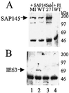
FIG. 1
FIG. 1
Coimmunoprecipitation of IE63 and SAP145 in vivo using antibodies directed against SAP145. HSV-1-infected (WT), 27-lacZ-infected, or mock-infected (MI) HeLa cell extracts were immunoprecipitated with SAP145 antiserum. Aliquots of the precipitated proteins were separated by SDS-polyacrylamide gel electrophoresis, transferred to nitrocellulose, and analyzed by Western blotting using SAP145 antiserum or IE63 monoclonal antibody. (A) Immunoprecipitates obtained with SAP145 antiserum (lanes 1 to 3) or preimmune (PI) serum (lane 4), Western blotted with SAP145 antiserum. (B) Immunoprecipitates obtained with SAP145 antiserum (lanes 1 to 3) or preimmune serum (lane 4), Western blotted with IE63 antiserum.
FIG. 2
FIG. 2
In vitro interaction between GST-IE63 and SAP145 and between GST-SAP145 and IE63. HSV-1-infected (WT) or mock-infected (MI) HeLa cell extracts were mixed with GST-IE63 (GST63), GST-SAP145 (GST145), or GST alone. Bound proteins were separated by SDS-polyacrylamide gel electrophoresis, transferred to nitrocellulose, and analyzed by Western blotting using SAP145 antiserum or IE63 monoclonal antibody. (A) Coomassie blue staining of the GST fusion proteins used in the pull-down assays. (B) Proteins binding to GST (lanes 3 and 4) or GST-SAP145 (lanes 5 and 6) following Western blotting with IE63 antiserum. The cell extracts used (lanes 1 and 2) were loaded with 25% of the amount used for the pull-downs. (C) Proteins binding to GST (lanes 1 and 2) or GST-IE63 (lanes 3 and 4), following Western blotting with SAP145 antiserum.
FIG. 3
FIG. 3
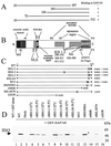
Mapping the interaction of IE63 with SAP145. (A) Truncations used in the yeast two-hybrid assay to map the IE63 region involved in interaction with SAP145; results of the interactions are shown as plus or minus signs. (B) Schematic representation (not to scale) of IE63 protein showing the different functional regions as described in reference . NES, leucine-rich nuclear export signal; NLS, nuclear localization signal; RGG box, arginine-rich RNA binding region; KH 1-3, hnRNP K homology domains; Sm, homology domain present in certain Sm proteins. (C) HSV-1 IE63 mutant viruses used to study the interaction with SAP145. X's indicate the locations of amino acid substitutions which are described on the right-hand side of the schematic; truncations also are shown, and the last amino acid is indicated. Mutation R480H in mutants BS3-3, BS4-3, BS5-3, and BSLG4 leads to a ts phenotype; the second mutations in these viruses are intragenic suppressors of this phenotype (see text). (D) Extracts from HeLa cells infected with viruses shown in panel C were mixed with GST alone (lane 1) or GST-SAP145 (lanes 2 to 16); bound proteins were separated by SDS-polyacrylamide gel electrophoresis, transferred to nitrocellulose, and analyzed by Western blotting using IE63 antiserum.
FIG. 4
FIG. 4
SAP145 interacts with hnRNP K and CK2 only in the presence of IE63. (A) HSV-1-infected (WT), 27-lacZ-infected (27), or mock-infected (MI) HeLa cell extracts were mixed with GST-hnRNP K (GST-K, lanes 1 to 3) or GST alone (lane 4). Bound proteins were separated by SDS-polyacrylamide gel electrophoresis, transferred to nitrocellulose, and analyzed by Western blotting using SAP145 antiserum. (B) Following immunoprecipitation with anti-SAP145 antiserum, as shown in Fig. 1, samples were analyzed for CK2 activity using a peptide assay. CK2 assays were performed with immunoprecipitates generated by preimmune (pi) serum (lane 3) and SAP145 (145) antiserum (lanes 1, 2, 4, and 5) from HSV-1 wt-infected (wt), 27-lacZ-infected (27lacZ), or mock-infected (mi) HeLa cell extracts and in the absence (lane 1) or presence (lanes 2 to 5) of peptide substrate.
FIG. 5
FIG. 5
IE63 colocalizes with SAP145 in HSV-1-infected cells. Immunofluorescence assay was performed on HeLa cells using anti-IE63 serum and anti-SAP145 serum. (A) HSV-1 wt virus-infected cells. (B) pCMV-63-transfected cells. (C) Mock-infected cells.
FIG. 6
FIG. 6
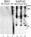
IE63 inhibits splicing in vitro and alters splicing complex formation. In vitro splicing assays were performed using adenovirus major late precursor mRNA with nuclear extracts from HSV-1 wt-infected (lanes 3), 27-lacZ-infected (lanes 2), or mock-infected (lanes 1) HeLa cells. The products of the splicing reaction were run on gels as follows. (A) A 10% polyacrylamide–8 M urea denaturing gel, to separate the RNA products formed. The pre-mRNA was 32P labeled, and products were visualized by exposure to X-ray film overnight. (B) A nondenaturing agarose-polyacrylamide composite gel to separate the protein-RNA complexes formed. Complexes bound to labeled RNA were visualized by exposure to X-ray film overnight.
FIG. 7
FIG. 7
IE63 and SAP145 proteins are found in splicing complex B/C. Duplicate samples of the protein complexes, as formed in the results shown in Fig. 6B, were transferred to nitrocellulose and assayed by Western blotting using SAP145 (lanes 4 to 6) or IE63 antiserum (lanes 1 to 3). Complexes were formed using HSV-1 wt-infected (WT), 27-lacZ-infected, or mock-infected (MI) HeLa cell nuclear extracts. The locations of the complexes as detected by autoradiography are indicated on the right-hand side.

References

    1. Ackermann M, Braun D K, Pereira L, Roizman B. Characterization of herpes simplex virus 1 α proteins 0, 4, and 27 with monoclonal antibodies. J Virol. 1984;52:108–118. - PMC - PubMed
    1. Baines J D, Roizman B. The cDNA of UL15, a highly conserved herpes simplex virus 1 gene, effectively replaces the two exons of the wild-type virus. J Virol. 1992;66:5621–5626. - PMC - PubMed
    1. Behrens S-E, Tyc K, Kastner B, Reichelt J, Lührmann R. Small nuclear ribonucleoprotein (RNP) U2 contains numerous additional proteins and has a bipartite RNP structure under splicing conditions. Mol Cell Biol. 1993;13:307–319. - PMC - PubMed
    1. Bryant H E, Matthews D A, Wadd S, Scott J E, Kean J, Graham S, Russell W C, Clements J B. Interaction between herpes simplex virus type 1 IE63 protein and cellular protein p32. J Virol. 2000;74:11322–11328. - PMC - PubMed
    1. Calvio C, Neubauer G, Mann M, Lamond A I. Identification of hnRNP P2 as TLS/FUS using electrospray mass spectrometry. RNA. 1995;1:724–733. - PMC - PubMed

Publication types

Substances

LinkOut - more resources