. 2001 May;75(9):4394-8.
doi: 10.1128/JVI.75.9.4394-4398.2001.

In vivo accumulation of cyclin A and cellular replication factors in autonomous parvovirus minute virus of mice-associated replication bodies

Affiliations

In vivo accumulation of cyclin A and cellular replication factors in autonomous parvovirus minute virus of mice-associated replication bodies

T Bashir et al. J Virol. 2001 May.

Abstract

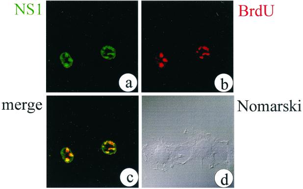
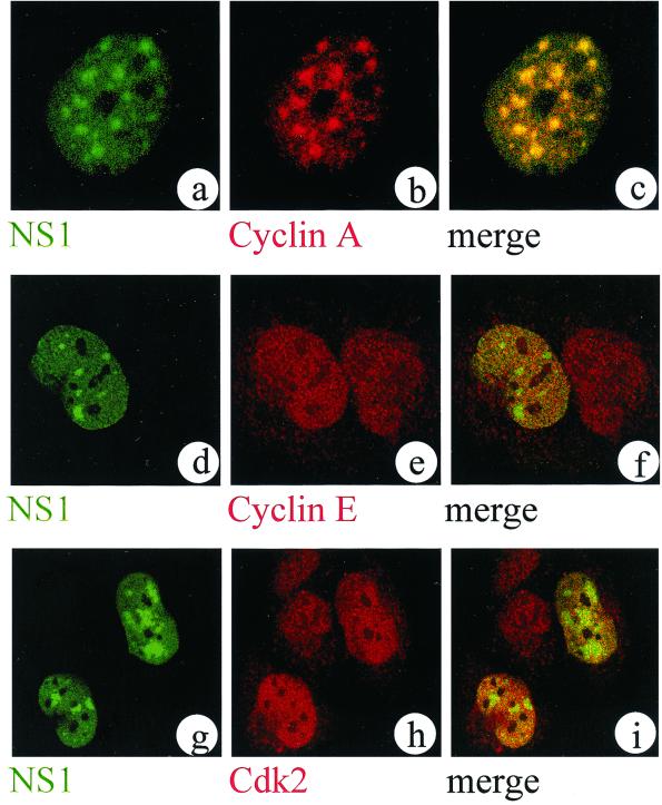
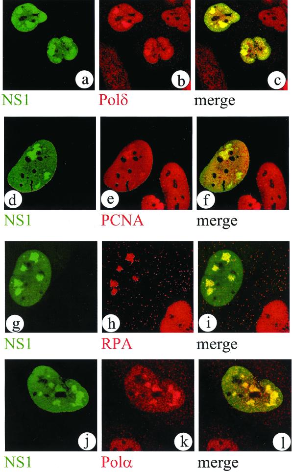
Autonomous parvovirus minute virus of mice (MVM) DNA replication is strictly dependent on cellular factors expressed during the S phase of the cell cycle. Here we report that MVM DNA replication proceeds in specific nuclear structures termed autonomous parvovirus-associated replication bodies, where components of the basic cellular replication machinery accumulate. The presence of DNA polymerases alpha and delta in these bodies suggests that MVM utilizes partially preformed cellular replication complexes for its replication. The recruitment of cyclin A points to a role for this cell cycle factor in MVM DNA replication beyond its involvement in activating the conversion of virion single-stranded DNA to the duplex replicative form.


Figures

FIG. 1
FIG. 1
MVM DNA replication colocalizes with NS1 in APAR bodies in the nuclei of infected A9 cells. Representative confocal optical sections through the nuclei of infected cells are shown. NS1 was localized with the SP8 polyclonal antiserum and a fluorescein isothiocyanate (FITC)-conjugated secondary antibody (a). Replication was monitored by incorporation of BrdU and indirect immunofluorescence using a BrdU-specific antibody and a tetramethyl rhodamine isothiocyanate (TRITC)-conjugated secondary antibody (b). In a merged image, colocalized structures from panels a and b appear yellow (c). By phase-contrast microscopy (Nomarski), the cells show no obvious sign of NS1-induced cytotoxicity at the time of fixation (15 h postinfection) (d).
FIG. 2
FIG. 2
Cyclin A, but not cyclin E or cdk2, accumulates in APAR bodies. Representative confocal images of nuclei from double-labeled MVM-infected A9 (a to c) or NBE (d to i) cells are shown. NS1 was detected with FITC using the NS1-specific 3D9 antibody (a generous gift from N. Salomé and D. Pintel) (a, d, and g). Images of cyclin A (b), cyclin E (e), and cdk2 (h) were detected with TRITC in the same confocal plane as shown in the left column using the respective primary antisera for cyclin A (a generous gift from M. Pagano), cyclin E (Ab-1; Neomarkers, Fremont, Calif.), and cdk2 (Ab-3; Neomarkers). The superimposition of both channels from the left and middle columns reveals either the colocalization (yellow signal [c]) or the lack of colocalization (distinct red and green signals [f and i]) of the respective factors in relationship to APAR bodies.
FIG. 3
FIG. 3
Cellular replication factors DNA polymerases δ and α, PCNA, and RPA accumulate in APAR bodies. Representative confocal images of nuclei from double-labeled MVM-infected NBE cells are shown. NS1 was detected with FITC using the SP8 antiserum (a, d, g, and j). Replication factors were detected with TRITC in the same confocal plane as shown in the left column, using the respective antibodies against DNA polymerase δ (Transduction Laboratories) (b), PCNA (PC10; Upstate Biotechnology) (e), RPA (Ab-1; Oncogene) (h), and DNA polymerase α (SJK132-20 [26]) (k), respectively. A merged image of both channels from the left and center columns provides evidence for colocalization of these factors in APAR bodies (c, f, i, and l).

References

    1. Bashir T, Horlein R, Rommelaere J, Willwand K. Cyclin A activates the DNA polymerase delta-dependent elongation machinery in vitro: a parvovirus DNA replication model. Proc Natl Acad Sci USA. 2000;97:5522–5527. - PMC - PubMed
    1. Brenot-Bosc F, Gupta S, Margolis R L, Fotedar R. Changes in the subcellular localization of replication initiation proteins and cell cycle proteins during G1-to S-phase transition in mammalian cells. Chromosoma. 1995;103:517–527. - PubMed
    1. Cardoso M C, Leonhardt H, Nadal-Ginard B. Reversal of terminal differentiation and control of DNA replication: cyclin A and Cdk2 specifically localize at subnuclear sites of DNA replication. Cell. 1993;74:979–992. - PubMed
    1. Christensen J. Binding of the major non-structural protein (NS1) of MVM to the large subunit of human replication protein is involved in origin unwinding. Infect Dis Rev. 2000;2:152.
    1. Christensen J, Cotmore S F, Tattersall P. A novel cellular site-specific DNA-binding protein cooperates with the viral NS1 polypeptide to initiate parvovirus DNA replication. J Virol. 1997;71:1405–1416. - PMC - PubMed

MeSH terms

LinkOut - more resources