. 2001 May;21(9):2991-3000.
doi: 10.1128/MCB.21.9.2991-3000.2001.

Identification of liver X receptor-retinoid X receptor as an activator of the sterol regulatory element-binding protein 1c gene promoter

Affiliations

Identification of liver X receptor-retinoid X receptor as an activator of the sterol regulatory element-binding protein 1c gene promoter

T Yoshikawa et al. Mol Cell Biol. 2001 May.

Abstract

In an attempt to identify transcription factors which activate sterol-regulatory element-binding protein 1c (SREBP-1c) transcription, we screened an expression cDNA library from adipose tissue of SREBP-1 knockout mice using a reporter gene containing the 2.6-kb mouse SREBP-1 gene promoter. We cloned and identified the oxysterol receptors liver X receptor (LXRalpha) and LXRbeta as strong activators of the mouse SREBP-1c promoter. In the transfection studies, expression of either LXRalpha or -beta activated the SREBP-1c promoter-luciferase gene in a dose-dependent manner. Deletion and mutation studies, as well as gel mobility shift assays, located an LXR response element complex consisting of two new LXR-binding motifs which showed high similarity to an LXR response element recently found in the ABC1 gene promoter, a reverse cholesterol transporter. Addition of an LXR ligand, 22(R)-hydroxycholesterol, increased the promoter activity. Coexpression of retinoid X receptor (RXR), a heterodimeric partner, and its ligand 9-cis-retinoic acid also synergistically activated the SREBP-1c promoter. In HepG2 cells, SREBP-1c mRNA and precursor protein levels were induced by treatment with 22(R)-hydroxycholesterol and 9-cis-retinoic acid, confirming that endogenous LXR-RXR activation can induce endogenous SREBP-1c expression. The activation of SREBP-1c by LXR is associated with a slight increase in nuclear SREBP-1c, resulting in activation of the gene for fatty acid synthase, one of its downstream genes, as measured by the luciferase assay. These data demonstrate that LXR-RXR can modify the expression of genes for lipogenic enzymes by regulating SREBP-1c expression, providing a novel link between fatty acid and cholesterol metabolism.


Figures

FIG. 1
FIG. 1
Screening of an SREBP-1 knockout mouse adipose tissue expression library to identify cDNA clones which activate the SREBP-1c promoter. An expression cDNA library from the adipose tissues of SREBP-1-null mice was constructed as described in Materials and Methods. A luciferase reporter gene containing the mouse SREBP-1c promoter (2.6 kb), pBP1c2600-Luc, and each subpool containing approximately 1,500 cDNA clones were cotransfected into HEK 293 cells with pSV-βgal as a reference plasmid. Luciferase activity was measured and normalized by β-galactosidase activity. After several rounds of screening, six positive single clones were isolated from the library (three LXRα and three LXRβ). A representative result from the initial screening is shown.
FIG. 2
FIG. 2
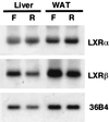
Northern blot analysis of LXRs from livers and white adipose tissues (WAT) from mice fasted and refed. Total RNA was prepared and pooled (10 μg) equally from either livers or adipose tissues of normal mice fasted (F) or refed (R) after fasting as described previously (41), blotted to a nylon membrane, and hybridized with the indicated cDNA probes. A cDNA probe for 36B4 (acidic ribosomal phosphoprotein P0) was used as a loading control.
FIG. 3
FIG. 3
Dose-dependent activation of SREBP-1c promoter by LXRs (left panel) and their ligands (right panel). (Left panel) pBP1c2600-Luc was cotransfected into HEK 293 cells with a positive clone expressing LXR from the screening (pCMV-LXRα or pCMV-LXRβ), an empty vector (CMV-7) used as a control, or pSV-βgal, which was used as a reference plasmid. (Right panel) Ligands for LXR or ethanol (used as a control) were added to the cells after transfection of pBP1c2600-Luc and pSV-βgal in medium with 10% fetal bovine serum 24 h prior to the assay. After the incubation, luciferase activity was measured and normalized to β-galactosidase activity. The fold induction of luciferase activity by LXRs or their ligands (means ± standard deviations in the left panel [n = 3]; means in the right panel [n = 2]) compared to that of controls is shown. Cho, cholesterol.
FIG. 4
FIG. 4
Activation of SREBP-1c promoter by LXR via the oxysterol-inducible region in the promoter. The previously identified oxysterol-inducible region and the SRE complex in the SREBP-1c promoter are illustrated at the top. pBP-1c2600-Luc and two shorter versions, pBP1c550- and pBP1c90-Luc, were prepared and used for transfection into 293 cells as described in Materials and Methods. Luciferase activity was measured and normalized to β-galactosidase activity. Fold induction of luciferase activity (mean ± standard deviation) by LXRs is shown.
FIG. 5
FIG. 5
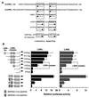
Identification of LXREs by deletional and mutational analysis of the oxysterol-inducible region in the SREBP1c-promoter. (A) DNA sequencing of the oxysterol-inducible region of the SREBP1c promoter identified two elements, which were designated mLXREa and -b. These elements are highly similar to the LXRE of the human ABC1 promoter. (B) Sequential deletion and mutation analysis of the oxysterol-inducible region was performed with reporter genes with the indicated sizes and positions of the promoter. HEK 293 cells were transfected with each reporter plasmid, pCMV-LXR, and reference plasmid pSV-βgal and thereafter cultured for 24 h in medium with 10% fetal bovine serum. Luciferase activity was measured and normalized to β-galactosidase activity. Fold induction of luciferase activity by LXRs (means ± standard deviations) is shown. SV40, simian virus 40.
FIG. 6
FIG. 6
Binding of LXR to LXREs in the SREBP1c promoter as measured by gel mobility shift assay. (A) LXREa and LXREb in the SREBP-1c promoter were labeled and incubated with nuclear extracts from transfected 293 cells in which LXRα or -β and RXR were overexpressed. A molar excess of unlabeled LXREa or -b fragment and antibody against LXRα and -β or RXRα were added to the incubations in competition and supershift assays, respectively. The DNA-protein complexes were resolved in a 4.8% polyacrylamide gel. The shifted and supershifted LXRE probes are indicated by thick and thin arrows, respectively. NR, nuclear extract from mock-transfected cells used as a negative control. (B) LXREa and -b in the oxysterol-inducible region of the SREBP-1c promoter are shown.
FIG. 7
FIG. 7
Dose-dependent activation of the LXRE complex enhancer in the SREBP-1c promoter by LXRs (left panel) and their ligands (right panel). The LXRE complex (LXREa and -b, bp −249 to −148) in the SREBP-1c promoter was fused to a luciferase reporter plasmid which contained a simian virus 40 promoter (pGL2 promoter vector; Fig. 5) to estimate LXRE enhancer activity (pLXRE-Luc). (Left panel) pLXRE-Luc was cotransfected into HEK 293 cells with pCMV-LXRα, pCMV-LXRβ, or an empty vector (CMV-7, used as a control) and pSV-βgal, which was used as a reference plasmid. (Right panel) Ligands for LXR or ethanol (used as a control) was added to the cells after transfection of pLXRE-Luc and pSV-βgal 24 h prior to the assay. After the incubation, luciferase activity was measured and normalized to β-galactosidase activity. Fold induction of luciferase activity by LXRs or their ligands (means ± standard deviations in the left panel [n = 3]; means in the right panel [n = 2]) compared to that of the control is shown. Cho, cholesterol.
FIG. 8
FIG. 8
Synergistic activation of the LXRE complex in the SREBP-1c promoter by LXRs, RXR, and their ligands. The luciferase reporter construct containing the LXRE complex in the mouse SREBP-1c promoter (pLXRE-Luc) was cotransfected into HEK 293 cells with pCMV-LXRα or -β, pRXR, or both and pSV-βgal. After the transfection, the cells were incubated in the absence or presence of the ligand 22(R)-HC, 9CRA, or both for 24 h. Luciferase activity was measured and normalized to β-galactosidase activity. Fold induction (means ± standard deviations) compared to that of the control (an empty expression vector with no addition of ligands) is shown.
FIG. 9
FIG. 9
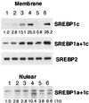
Induction of SREBP-1c protein by LXR-RXR ligands in HepG2 cells as measured by immunoblot analysis. HepG2 cells were incubated with the indicated ligands for LXR or RXR for 24 h. In some groups, 1 μg of 25-HC per ml and 10 μg of cholesterol per ml were also added to the medium to suppress sterol-regulatory cleavage activity of SREBPs (sterol-suppressed condition). After the incubation, nuclear extracts and membranes were prepared from the cells and aliquots (30 μg) of protein were subjected to immunoblot analysis using antibodies against SREBP-1c, SREBP-1a and -1c, and SREBP-2. The bands were visualized with the ECL system. The values below the bands refer to relative fold changes in protein levels. Lanes: 1, ethanol; 2, 22(R)-HC; 3, 9CRA; 4, 22(R)-HC plus 9CRA; 5, ethanol in a sterol-suppressed condition; 6, 22(R)-HC plus 9CRA in a sterol-suppressed condition.
FIG. 10
FIG. 10
SREBP-1, -2, and FAS mRNA levels in HepG2 cells treated with LXR or RXR ligands as measured by Northern blot analysis. HepG2 cells were incubated with the indicated ligands for LXR or RXR for 24 h. In some groups, 25-HC at 1 μg/ml and cholesterol at 10 μg/ml were added to the medium to suppress the sterol-regulatory cleavage activity of SREBPs (sterol-suppressed condition). Total RNA was prepared and pooled (10 μg) equally from HepG2 cells, blotted to a nylon membrane, and then hybridized with the indicated cDNA probes. The values below the bands refer to relative fold changes in mRNA levels normalized relative to the 36B4 signal. Lanes: 1, ethanol; 2, 22(R)-HC; 3, 9CRA; 4, 22(R)-HC plus 9CRA; 5, ethanol in a sterol-suppressed condition; 6, 22(R)-HC plus 9CRA in a sterol-suppressed condition. A cDNA probe for 36B4 (acidic ribosomal phosphoprotein P0) was used as a loading control.
FIG. 11
FIG. 11
Activation of the FAS promoter by LXRs and their ligands. A luciferase reporter construct containing the rat FAS promoter (31) and reference plasmid pSV-βgal with empty plasmid pCMV7 or expression plasmids pCMV-LXR and pRXR were transfected into HepG2 cells. After the transfection, ligands for LXR and RXR or ethanol (used as a negative control) was added to the cells, followed by a 24-h incubation in the medium with 10% fetal bovine serum. In some sets, 25-HC at 1 μg/ml and cholesterol at 10 μg/ml were also added to the medium to suppress the sterol-regulatory cleavage activity of SREBPs (sterol-suppressed condition). Luciferase activity was measured and normalized to β-galactosidase activity. Fold induction (means ± standard deviations) by LXRs is shown. pCMV-nSREBP-1c, an expression plasmid of human nuclear SREBP-1c was used as a positive control for FAS promoter activation (BP-1c). Lα+R, LXRα plus RXR; 22+9, 22(R)-HC plus 9CRA.
FIG. 12
FIG. 12
Dual regulation of SREBP-1c promoter by LXR-RXR and SREBPs. In the presence of excess amount of sterols, presumably a concomitant increase in oxysterol levels activates the SREBP-1c promoter through the LXRE complex while the cleavage activity of SREBPs is suppressed (A). When cells are deprived of sterols, increased nuclear SREBPs activate the SREBP-1c promoter through the SRE complex (B). The dual activation of the SREBP-1c promoter through LXRE and SRE complexes ensures the maintenance of lipogenesis irrespective of cellular sterol levels.

Similar articles

Cited by

References

    1. Amemiya-Kudo M, Shimano H, Yoshikawa T, Yahagi N, Hasty A H, Okazaki H, Tamura Y, Shionoiri F, Iizuka Y, Ohashi K, Osuga J-I, Harada K, Gotoda T, Sato R, Kimura S, Ishibashi S, Yamada N. Promoter analysis of the mouse sterol regulatory element-binding protein (SREBP)-1c gene. J Biol Chem. 2000;275:31078–31085. - PubMed
    1. Andersson S, Davis D L, Dahlback H, Jornvall H, Russell D W. Cloning, structure, and expression of the mitochondrial cytochrome P-450 sterol 26-hydroxylase, a bile acid biosynthetic enzyme. J Biol Chem. 1989;264:8222–8229. - PubMed
    1. Brown M S, Goldstein J L. A proteolytic pathway that controls the cholesterol content of membranes, cells, and blood. Proc Natl Acad Sci USA. 1999;96:11041–11048. - PMC - PubMed
    1. Brown M S, Goldstein J L. The SREBP pathway: regulation of cholesterol metabolism by proteolysis of a membrane-bound transcription factor. Cell. 1997;89:331–340. - PubMed
    1. Brown M S, Ye J, Rawson R B, Goldstein J L. Regulated intramembrane proteolysis: a control mechanism conserved from bacteria to humans. Cell. 2000;100:391–398. - PubMed

Publication types

MeSH terms

LinkOut - more resources