. 2001 May;21(9):3025-36.
doi: 10.1128/MCB.21.9.3025-3036.2001.

Caspase cleavage enhances the apoptosis-inducing effects of BAD

Affiliations

Caspase cleavage enhances the apoptosis-inducing effects of BAD

F Condorelli et al. Mol Cell Biol. 2001 May.

Abstract

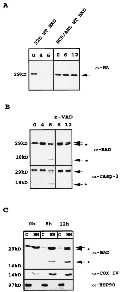
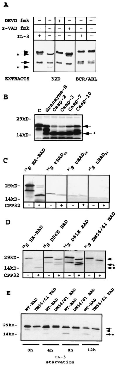
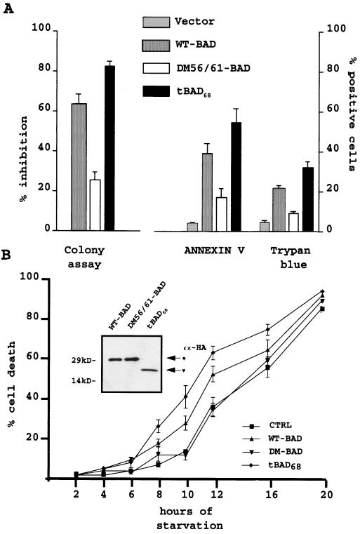
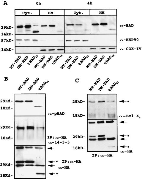
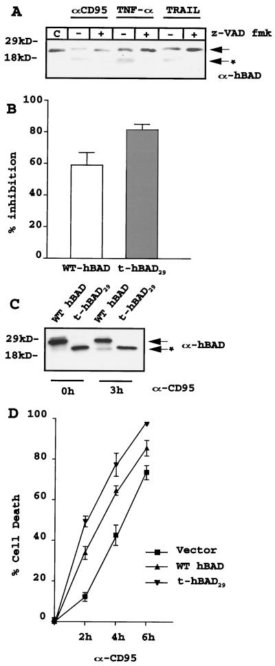
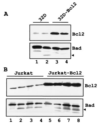
The function of BAD, a proapoptotic member of the Bcl-2 family, is regulated primarily by rapid changes in phosphorylation that modulate its protein-protein interactions and subcellular localization. We show here that, during interleukin-3 (IL-3) deprivation-induced apoptosis of 32Dcl3 murine myeloid precursor cells, BAD is cleaved by a caspase(s) at its N terminus to generate a 15-kDa truncated protein. The 15-kDa truncated BAD is a more potent inducer of apoptosis than the wild-type protein, whereas a mutant BAD resistant to caspase 3 cleavage is a weak apoptosis inducer. Truncated BAD is detectable only in the mitochondrial fraction, interacts with BCL-X(L) at least as effectively as the wild-type protein, and is more potent than wild-type BAD in inducing cytochrome c release. Human BAD, which is 43 amino acids shorter than its mouse counterpart, is also cleaved by a caspase(s) upon exposure of Jurkat T cells to anti-FAS antibody, tumor necrosis factor alpha (TNF-alpha), or TRAIL. Moreover, a truncated form of human BAD lacking the N-terminal 28 amino acids is more potent than wild-type BAD in inducing apoptosis. The generation of truncated BAD was blocked by Bcl-2 in IL-3-deprived 32Dcl3 cells but not in Jurkat T cells exposed to anti-FAS antibody, TNF-alpha, or TRAIL. Together, these findings point to a novel and important role for BAD in maintaining the apoptotic phenotype in response to various apoptosis inducers.


Figures

FIG. 1
FIG. 1
(A) Expression of WT HA-BAD in IL-3-starved parental and BCR/ABL-expressing 32Dcl3 cells. Extracts from cells starved for 0, 4, and 6 h (32D) or 0, 6, and 12 h (BCR/ABL) were blotted with anti-HA antibody (α-HA). (B) Effect of z-VAD fmk (50 μM) on levels of WT HA-BAD in IL-3-starved parental 32Dcl3 cells. Western blot analyses were performed with an anti-BAD (C-20, upper gels) antibody. Caspase-3 levels were measured as a control for the inhibitory capacity of z-VAD fmk. Results are representative of three separate experiments. (C) HA-BAD expression in subcellular fractions from retrovirus-infected parental cells. At 0, 8, and 12 h after IL-3 withdrawal, mitochondrial (HM) and cytoplasmic (C) extracts were tested for BAD expression using the anti-BAD (C-20) antibody. Arrows indicate full-length HA-BAD; arrows plus asterisks indicate cleaved products of BAD or caspase 3. Results are representative of three separate experiments.
FIG. 2
FIG. 2
(A) Cleavage of in vitro-translated 35S-labeled murine HA-BAD by extracts of IL-3-starved (12 h) parental cells, untreated or treated with z-VAD fmk (50 μM) or DEVD fmk (50 nM), and nonstarved or starved (12 h) BCR/ABL-expressing 32Dcl3 cells. (B) Cleavage of in vitro-translated 35S-labeled murine BAD by recombinant granzyme-B, caspase 2, caspase 3, caspase 7, and caspase 10. Lane C contains the uncleaved in vitro translation products. (C and D) caspase 3-dependent cleavage of in vitro-translated 35S-labeled full-length, truncated, and mutant forms of murine HA-BAD. Reactions were carried out for 1 h at 30°C and stopped by adding sample buffer. The down-shift of full-length BAD produced by caspase 3 on WT and mutated BAD is probably due to the cleavage of the HA epitope, which contains a caspase 3 canonical cleavage site (DEVD). (E) Kinetics of WT and DM56/61 BAD-HA expression in virus-infected 32Dcl3 cells 0, 4, 8, and 12 h after IL-3 removal. Anti-BAD (C-20) antibody was used for Western blotting. Arrows indicate full-length BAD and a shorter translation product (∼23 kDa), presumably starting from methionine 43 of the full-length coding sequence; arrows plus asterisks indicate the cleavage products.
FIG. 3
FIG. 3
(A) Colony formation, ANNEXIN V reactivity, and trypan blue staining in 32Dcl3 cells transiently overexpressing WT, double-mutant (DM56/61), or truncated (tBAD68) BAD. For clonogenic assays, parental 32Dcl3 cells were cultured in pLXSP, pLXSP WT BAD-HA pLXSP DM56/61 BAD-HA, or pLXSP tBAD68 BAD-HA retrovirus-containing medium. Twelve hours after infection, cells were plated in methylcellulose in the presence of IL-3 and puromycin (2.5 μg/ml). Colonies were counted 5 days later. The effect of BAD (WT, mutant, and truncated) on the clonogenic ability of infected cells is expressed as the percentage (mean + standard deviation [SD]) of inhibition of the clonogenic ability of vector-infected cells. For ANNEXIN V reactivity or trypan blue uptake, 32Dcl3 cells were cultured with the supernatant from pMIGR1-WT BAD, pMIGR1-DM56/61 HA-BAD, or pMIGR1-tBAD68 HA-BAD retrovirus-infected PHOENIX cells. At 12 h after infection, GFP-positive cells were sorted by flow cytometry. Transiently infected cells were probed with ANNEXIN V 2 h later. Data are expressed as percentages (means + SDs) of ANNEXIN V-positive cells versus total viable cells (7ADD negative). 32Dcl3 GFP-positive cells were also stained with trypan blue and counted. Results are expressed as the percentages (means + SDs) of trypan blue-positive cells among total GFP-positive cells. Five hundred cells were counted in each experiment. Results are representative of four separate experiments. (B) Kinetics of cell death in IL-3-starved parental 32Dcl3 cells (CTRL) and 32Dcl3 cells stably expressing WT, DM56/61, or tBAD68 HA-BAD. Cell death was measured as a percentage (mean + SD) of trypan blue-positive cells among total cells. Five hundred cells were counted in each experiment. Results are representative of three separate experiments. The inset shows a Western blot of WT, BAD, DM56/61 BAD, and tBAD68 expression in the 32Dc13-infected cells used in the cell death assay. Anti-HA antibody (α-HA) was used as the probe.
FIG. 4
FIG. 4
(A) BAD (WT, mutant, and truncated) expression in subcellular fractions of retrovirus-infected parental 32Dc13 cells. Western blots show BAD levels in mitochondrion (HM)- and cytoplasm (Cyt)-enriched fractions from parental cells in the presence of IL-3 or 4 h after its removal (upper gels). Anti-BAD (C-20) antibody (α-BAD) was used as the probe. Levels of the cytoplasmic marker HSP90 (middle gels) and mitochondrial marker subunit IV of cytochrome oxidase (COX-IV, lower gels) were measured as a control for equal loadings and the purity of subcellular fractions. Results are representative of three separate experiments. (B) Serine phosphorylation and 14-3-3 interaction of WT, DM56/61, and tBAD68 HA-BAD in retrovirus-infected parental cells. Western blot analyses were carried out with a mix of anti-pSer112 and -pSer136 antibodies to monitor phosphorylated BAD (upper gel). To study the interaction between 14-3-3 protein and WT, DM56/61, and tBAD68 HA-BAD, cell extracts were immunoprecipitated with the anti-HA antibody and blotted with anti-14-3-3 antibody (middle gel). Levels of WT, DM56/61, and tBAD68 HA-BAD were measured as a control for equal loadings (lower gel). Results are representative of three separate experiments. Arrows indicate heavy- and light-chain immunoglobulins; arrows with asterisks indicate antibody-specific reactive bands. IP, immunoprecipitate. (C) Interaction of HA-BAD (WT, mutant, and truncated) with BCL-XL in retrovirus-infected 32Dc13 cells. Anti-HA immunoprecipitates from WT, DM56/61, and tBAD68 HA-BAD retrovirus-infected 32Dc13 cells were blotted with anti-BCL-XL antibody (upper gel). Levels of HA-tagged protein were measured as a control for immunoprecipitation (lower gel). Results are representative of three separate experiments. Arrows indicate heavy- and light-chain immunoglobulins; arrows with asterisks indicate antibody-specific reactive bands.
FIG. 5
FIG. 5
(A) Mitochondrial cytochrome c release induced by recombinant WT, DM56/61, and tBAD68 BAD proteins. Recombinant WT, double-mutant, and truncated BAD proteins fused to the TAT leader sequence were generated and purified as described in Materials and Methods. Partially purified mitochondria (15 μl) from parental 32Dc13 cells were incubated with a vehicle (lane 1; 250 mM sucrose buffer) or 100 nM WT BAD (lanes 2 and 3), double-mutant BAD (lanes 4 and 5), or truncated BAD (lane 6) in the absence (lanes 2, 4, and 6) or presence (lanes 1, 3, and 5) of 1 μl of recombinant caspase 3 (10 ng) in 30 μl. After 1 h at 30°C, samples were centrifuged at 12,000 × g for 10 min at 4°C. Supernatants (Sup) were subjected to an SDS–4 to 15% gradient polyacrylamide gel and transferred to a nitrocellulose filter. The filter was probed with a monoclonal anti-cytochrome c antibody (α-Cytochrome C) (upper gel). Mitochondrial pellets were lysed, and protein extracts were blotted with an antibody to subunit IV of cytochrome oxidase COX-IV) as a control for equal loadings (middle gel). The same filter was blotted with anti-cytochrome c antibody to monitor the presence of residual protein in the mitochondrial fraction (lower gel). (B) Densitometric analysis of cytochrome c release. The intensities of the cytochrome c band in the supernatants were measured and normalized against that of COX-IV detected in the pellet extracts, used as control (CTRL) of mitochondrial loading. Data are the ratios of the value of each cytochrome c band to that of its COX-IV counterpart. Results are representative of two separate experiments.
FIG. 6
FIG. 6
(A) Caspase-dependent cleavage of endogenous BAD in human Jurkat T cells after treatment with anti-Fas antibody (αCD95; 0.05 ng/ml), TNF-α (10 ng/ml), or TRAIL (1 μg/ml). Cells (2 × 10⁵/ml) were exposed to anti-CD95 antibody, TNF-α, or TRAIL for 6 h in the absence or presence of the caspase inhibitor z-VAD fmk (50 μM) and harvested. Cell extracts were subjected to an SDS–4 to 15% gradient polyacrylamide gel, transferred to a nitrocellulose membrane, and blotted with anti-BAD polyclonal rabbit antibody. Untreated cell extracts were used as a negative control for caspase activity. Results are representative of two separate experiments. The arrow indicates full-length hBAD; the arrow plus the asterisk indicates cleavage products. (B) Inhibition of colony formation by transient overexpression of WT or truncated hBAD. Parental Jurkat T cells were incubated with infectious supernatant from pLXSP, pLXSP WT hBAD, or pLXSP t-hBAD29 retrovirus-infected cells. After three cycles of infection (see Materials and Methods), cells were plated in methylcellulose in the presence of puromycin (2.5 μg/ml). Colonies were counted 7 days later. The effect of BAD on the clonogenic capacity of infected cells is expressed as a percentage (mean + SD) of inhibition of the clonogenic ability of vector-infected cells. Results are representative of three separate experiments. (C) Expression of exogenous WT and t-hBAD29 in infected-Jurkat T cells. A Western blot shows expression of WT and t-hBAD29 in a representative clone of Jurkat T cells, untreated or treated (3 h) with 0.05 ng of anti-CD95 antibody per ml. Protein extracts were blotted with a polyclonal anti-hBAD serum (H-168). Results are representative of two separate experiments. The arrow indicates full-length hBAD; the arrow with the asterisk indicates the cleavage product. (D) Kinetics of apoptosis in Jurkat T cells (empty vector or BAD transduced) treated with the anti-CD95 antibody for 0, 2, 4, and 6 h. Cell death was measured as a percentage (mean + SD) of trypan blue-positive cells among total cells. Results are representative of three separate experiments.
FIG. 7
FIG. 7
(A) Ectopic Bcl-2 expression blocks BAD cleavage in IL-3-starved 32Dc13 cells. Parental (lanes 1 and 2) and Bcl-2-expressing (lanes 3 and 4) 32Dc13 cells were infected with pLXSP-HA WT BAD; after selection in puromycin (2.5 μg/ml), transduced cells (2 × 10⁵/ml) expressing high levels of murine BAD were deprived of IL-3 for 8 h (lanes 2 and 4) and harvested. Cell extracts were subjected to an SDS–4 to 15% gradient polyacrylamide gel, transferred to a nitrocellulose membrane, and blotted with the anti-C-terminus (C-20) BAD antibody. (B) Ectopic Bcl-2 expression does not block BAD cleavage in death ligand-treated Jurkat T cells. Parental (lanes 1 to 4) and Bcl-2-expressing (lanes 5 to 8) Jurkat cells were infected with pLXSP WT hBAD; after selection in puromycin (2.5 μg/ml), transduced cells (2 × 10⁵/ml) expressing high levels of hBAD were treated for 6 h with anti-FAS antibody (lanes 2 and 6), TNF-α (lanes 3 and 7), and TRAIL (lanes 4 and 8) and harvested. Cell extracts were subjected to an SDS–4 to 15% gradient polyacrylamide gel, transferred to a nitrocellulose membrane, and blotted with polyclonal anti-BAD serum (H-168). Arrows indicate cleaved BAD.

References

    1. Adams J M, Cory S. The Bc1–2 protein family: arbiters of cell survival. Science. 1998;281:1322–1326. - PubMed
    1. Ayllon V, Martinez-A C, Garcia A, Cayla X, Rebollo A. Protein phosphatase 1alpha is a Ras-activated BAD phosphatase that regulates interleukin-2 deprivation-induced apoptosis. EMBO J. 2000;19:2237–2246. - PMC - PubMed
    1. Barry M, Heibein J A, Pinkoski M J, Lee S-F, Moyer R W, Green D R, Bleackley R C. Granzyme B short-circuits the need for caspase-8 activity during granule-mediated cytotoxic T-lymphocyte killing by directly clearing Bid. Mol Cell Biol. 2000;20:3781–3794. - PMC - PubMed
    1. Chandler J M, Cohen G M, MacFarlane M. Different subcellular distribution of caspase-3 and caspase-7 following Fas-induced apoptosis in mouse liver. J Biol Chem. 1998;273:10815–10818. - PubMed
    1. Chen Q, Gong B, Almasan A. Distinct stages of cytochrome c release from mitochondria: evidence for a feedback amplification loop linking caspase activation to mitochondrial dysfunction in genotoxic stress-induced apoptosis. Cell Death Differ. 2000;7:227–233. - PMC - PubMed

Publication types

MeSH terms