. 2001 May;21(9):3071-82.
doi: 10.1128/MCB.21.9.3071-3082.2001.

Negative regulation of CD4 gene expression by a HES-1-c-Myb complex

Affiliations

Negative regulation of CD4 gene expression by a HES-1-c-Myb complex

R D Allen 3rd et al. Mol Cell Biol. 2001 May.

Abstract

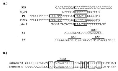
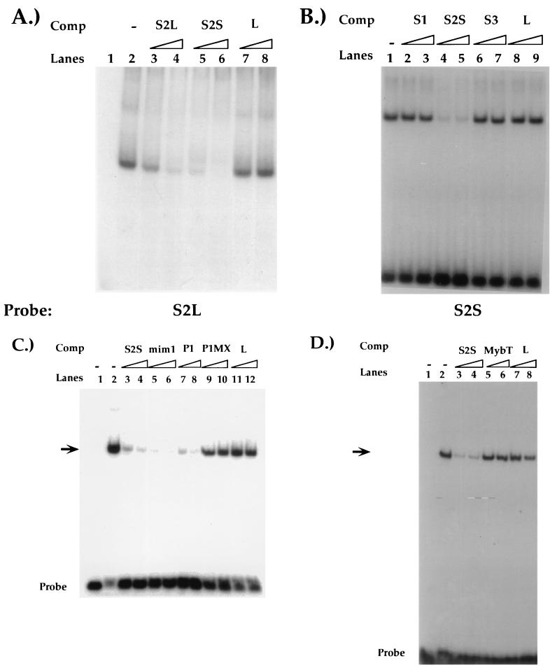
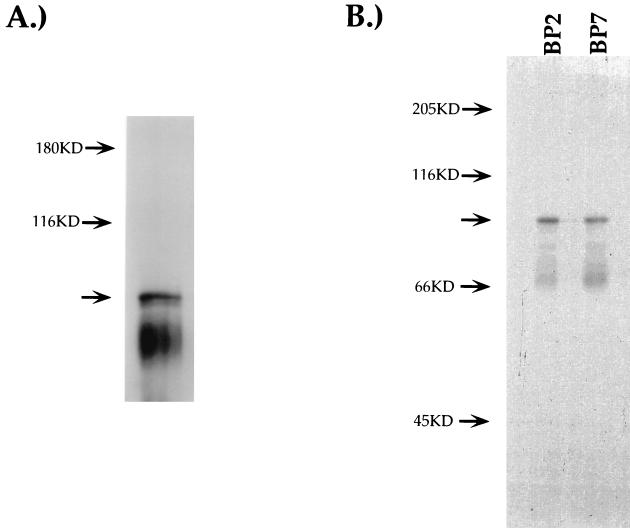
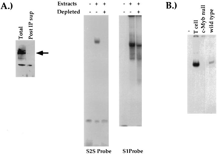
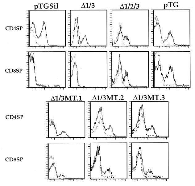
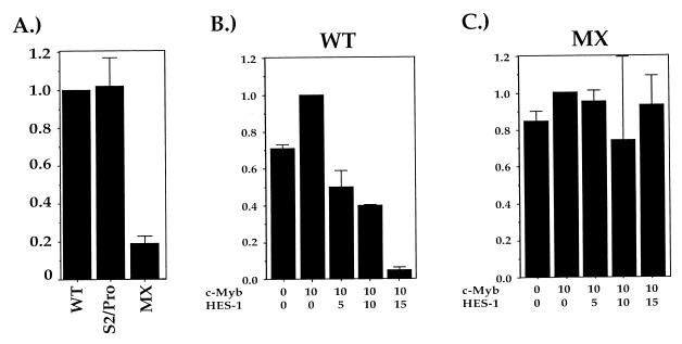
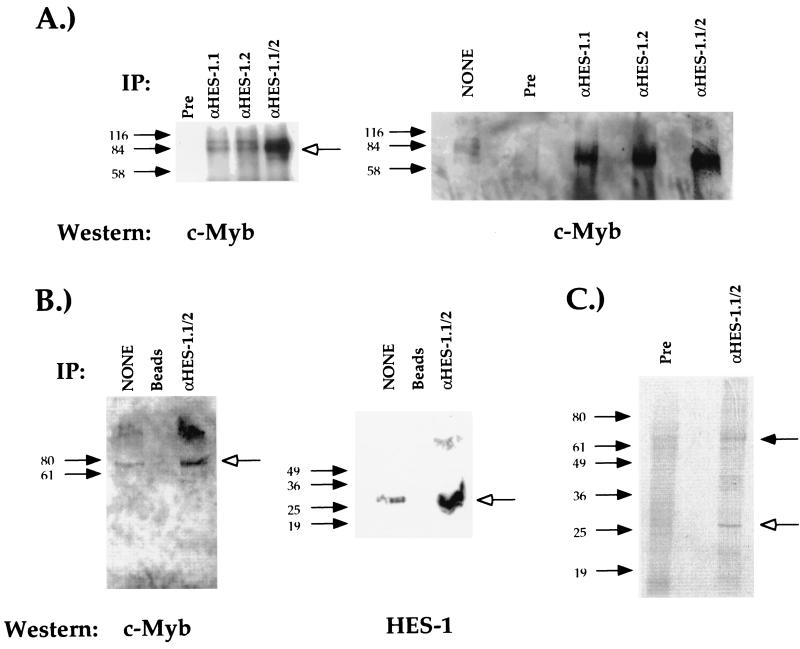
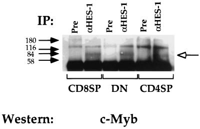
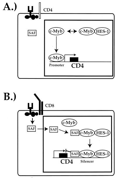
Expression of the CD4 gene is tightly controlled throughout thymopoiesis. The downregulation of CD4 gene expression in CD4(-) CD8(-) and CD4(-) CD8(+) T lymphocytes is controlled by a transcriptional silencer located in the first intron of the CD4 locus. Here, we determine that the c-Myb transcription factor binds to a functional site in the CD4 silencer. As c-Myb is also required for CD4 promoter function, these data indicate that depending on the context, c-Myb plays both positive and negative roles in the control of CD4 gene expression. Interestingly, a second CD4 silencer-binding factor, HES-1, binds to c-Myb in vivo and induces it to become a transcriptional repressor. We propose that the recruitment of HES-1 and c-Myb to the silencer leads to the formation of a multifactor complex that induces silencer function and repression of CD4 gene expression.


Figures

FIG. 1
FIG. 1
(A) Sequences of oligonucleotides used in the competition EMSA analyses. Boxed regions indicate consensus c-Myb recognition sequences within each oligonucleotide. The HES-1 and SAF DNA recognition sequences in S1 and S3, respectively, are overlined. (B) The DNA sequence identities between the P1 and S2 regions are indicated by boxed nucleotides. Consensus c-Myb recognition sequences are overlined.
FIG. 2
FIG. 2
(A) Characterization of the S2-binding factor. EMSAs using a 3′-extended radioactive S2L probe and CD4 SP TH D10 nuclear extracts. Reactions were performed in the absence of competitor (Comp) oligonucleotides (lane 2) or in the presence of excess S2L (lanes 3 and 4), S2S (lanes 5 and 6), or nonspecific (L, lanes 7 and 8) oligonucleotides. Lane 1, probe only. (B) EMSAs using the radioactive S2 probe and CD4 SP TH D10 nuclear extracts. Reactions were performed in the absence of competitor oligonucleotides (lane 1) or in the presence of excess S1 (lanes 2 and 3), S2S (lanes 4 and 5), S3 (lanes 6 and 7), or nonspecific (L, lanes 8 and 9) oligonucleotides. Sequences of the S2S probe and competitors are listed in Fig. 1. (C) Reactions were performed in the absence of competitor oligonucleotides (lane 2) or in the presence of excess S2S (lanes 3 and 4), mim-1 (lanes 5 and 6), P1 (lanes 7 and 8), P1MX (lanes 9 and 10), or nonspecific (L, lanes 11 and 12) oligonucleotides. Lane 1, probe only. (D) Reactions were performed in the absence of competitor oligonucleotides (lane 2) or in the presence of excess S2S (lanes 3 and 4), MybT (lanes 5 and 6), or nonspecific (L, lanes 7 and 8) oligonucleotides. Unlabeled oligonucleotides were used at 100- and 300-fold molar excesses. Sequences of competitor oligonucleotides are shown in Fig. 1. Arrows indicate S2-specific binding complex; free probe is indicated. Lane 1, probe only.
FIG. 3
FIG. 3
c-Myb binding to S2. EMSA reactions using a radioactive S2S probe and CD4 SP TH D10 nuclear extracts. (A) EMSA reactions were exposed to UV light for 15 min, resolved on SDS–10% PAGE gels, and visualized by autoradiography. (B) EMSA reactions were treated with UV light as for panel A and subsequently were immunoprecipitated using either the BP2 or BP7 anti-Myb antiserum. Immunoprecipitates were resolved on SDS–10% PAGE gels and visualized by autoradiography. Arrows indicate putative c-Myb DNA complexes.
FIG. 4
FIG. 4
c-Myb is necessary for formation of S2-binding complex. (A) Western blot of AKR1G1 DP nuclear extracts either untreated or depleted of c-Myb (left panel). The arrow indicates c-Myb. AKR1G1 DP nuclear extracts were either mock-treated or depleted of c-Myb and used in EMSA reactions with either the S2S (center panel) or the S1 (right panel) radioactive probes. (B) EMSA reactions using a radiolabeled S2 probe and extracts from the CD4 SP TH D10 clone, c-Myb null ES cells, or wild-type ES cells. See Materials and Methods for details. Post IP sup, postimmunoprecipitation supernatant.
FIG. 5
FIG. 5
The c-Myb-binding site in S2 is critical for CD4 silencer function. CD8 SP and CD4 SP T cells from pTG, pTGΔ1Δ3, pTGΔ1Δ2Δ3, pTGMT, or pTG transgenic mouse lines were gated on and analyzed for HLA-B7 expression. The presence of CD8+ HLA-B7+ T cells in the pTG, pTGΔ1Δ2Δ3, and pTGMT mice indicates a loss of silencer function in these constructs. Solid and dashed lines indicate staining with the anti-marker antibody and the isotype-matched control, respectively. Multiple founders for each construct were generated and analyzed; typical results are shown.
FIG. 6
FIG. 6
HES-1 modifies c-Myb function. (A) The c-Myb sites in the promoter and silencer are functionally interchangeable. Luciferase constructs containing the CD4 promoter with the c-Myb recognition site either intact (WT), mutated (MX), or substituted with the silencer c-Myb site (S2/Pro) were transfected into CD4 SP TH-clone D10 cells, and extracts from these cells were assayed for luciferase activity. Bars indicate percent of promoter activity when compared to that of the wild-type minimal CD4 promoter (100%). Data shown are compiled from at least three independent experiments with each construct. (B and C) HES-1 induces c-Myb to become a transcriptional repressor. The D10 CD4 SP TH clone was transfected with luciferase reporter constructs containing the CD4 promoter with either the c-Myb sites intact (B) or mutated (the MX mutation; panel C), a c-Myb expression vector, and increasing amounts of a HES-1 expression vector; error bars represent one standard deviation. Data are presented as fractions of the values obtained with the reporter construct and the c-Myb expression vector alone; typical values are 2 × 10⁴ to 4 × 10⁴ light units for the WT construct and 1 × 10³ to 3 × 10³ for the MX construct.
FIG. 7
FIG. 7
c-Myb binds to HES-1 in vivo. (A) Nuclear extracts from the AKR1G1 DP thymoma were immunoprecipitated (IP) with either the anti-HES-1 (α-HES-1) or preimmune (Pre) serum, resolved on an SDS-PAGE gel, and blotted with an anti-c-Myb antibody. Open arrows indicate the position of specific complex. Two different antisera against HES-1 generated from two different rabbits were tested either separately (α-HES-1.1 and α-HES-1.2) or pooled (α-HES-1.1/2). The lane marked “none” in the left panel represents a direct loading of 20% of the nuclear extract used in the immunoprecipitations. Densitometric analyses (see Materials and Methods) indicate that the input band is 6.6× less intense than the c-Myb bands in the IP lanes, indicating that 35% of the c-Myb in the nuclear extract is being immunoprecipitated with the HES-1 antiserum. (B) Nuclear extracts from the AKR1G1 DP thymoma were immunoprecipitated using either the HES-1 pooled antisera coupled to Sepharose beads (α-HES-1.1/2) or beads alone (beads), loaded onto an SDS-PAGE gel, and blotted with either the antibody against c-Myb (left panel) or the pooled HES-1 antisera (right panel). The lanes marked “none” in the left and right panels represent a direct loading of 20 and 10% of the nuclear extract used in the immunoprecipitations, respectively. (C) Direct immunoprecipitation of both c-Myb and HES-1 with the pooled HES-1 antisera. The S49 T-cell lymphoma was grown in 35S-methionine, and whole-cell extracts were purified as described previously (3). The labeled extracts were then immunoprecipitated either with the pooled HES-1 antisera (α-HES-1.1/2) or with the pooled preimmune sera (Pre), and the immunoprecipitates were resolved on an SDS-PAGE gel and visualized by autoradiography. Filled and open arrows indicate protein species of the molecular masses of c-Myb and HES-1, respectively.
FIG. 8
FIG. 8
HES-1 and c-Myb interact in all T cells. In vivo immunoprecipitations (IP) using anti-HES-1 (α-HES-1) or preimmune (Pre) serum with the D10 CD4 SP TH clone (CD4SP), the L3 CD8 SP TC clone (CD8SP), and the S49 DN thymoma and the pooled antisera. The open arrow indicates protein species of the molecular weight of c-Myb.
FIG. 9
FIG. 9
A model for the mechanism of CD4 gene expression control by c-Myb and HES-1. (A) Induction of CD4 promoter function by c-Myb during CD4 SP TH-cell development. (B) Induction of CD4 silencer function by a HES-1–c-Myb complex during CD8 SP TC-cell development. See the text for details.

Similar articles

Cited by

References

    1. Adlam M, Duncan D D, Ng D K, Siu G. Positive selection induces CD4 promoter and enhancer function. Int Immunol. 1997;9:877–887. - PubMed
    1. Allen R D, Bender T P, Siu G. c-Myb is essential for early T cell development. Genes Dev. 1999;13:1073–1078. - PMC - PubMed
    1. Ausubel F M, Brent R, Kingston R E, Moore D D, Seidman J G, Smith J A, Struhl K. Current protocols in molecular biology. New York, N.Y: John Wiley & Sons, Inc.; 1995.
    1. Badiani P, Corbella P, Kioussis D, Marvel J, Weston K. Dominant interfering alleles define a role for c-Myb in T-cell development. Genes Dev. 1994;8:770–782. - PubMed
    1. Berg L J, Pullen A M, Fazekas de St. Groth B, Mathis D, Benoist C, Davis M M. Antigen/MHC-specific T cells are preferentially exported from the thymus in the presence of their MHC ligand. Cell. 1989;58:1035–1046. - PubMed

Publication types

MeSH terms