. 2001 May;21(9):3126-36.
doi: 10.1128/MCB.21.9.3126-3136.2001.

Vav-Rac1-mediated activation of the c-Jun N-terminal kinase/c-Jun/AP-1 pathway plays a major role in stimulation of the distal NFAT site in the interleukin-2 gene promoter

Affiliations

Vav-Rac1-mediated activation of the c-Jun N-terminal kinase/c-Jun/AP-1 pathway plays a major role in stimulation of the distal NFAT site in the interleukin-2 gene promoter

O Kaminuma et al. Mol Cell Biol. 2001 May.

Abstract

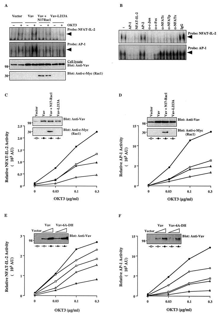
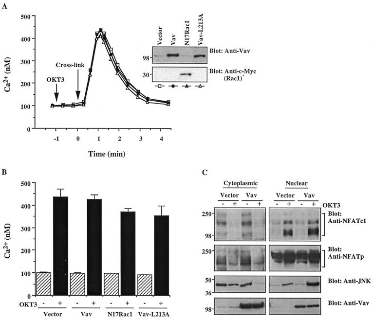
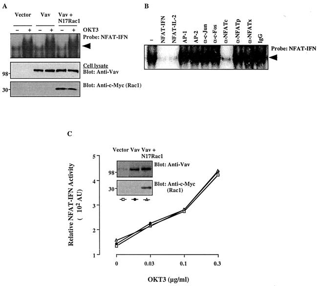
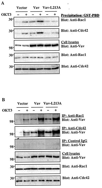
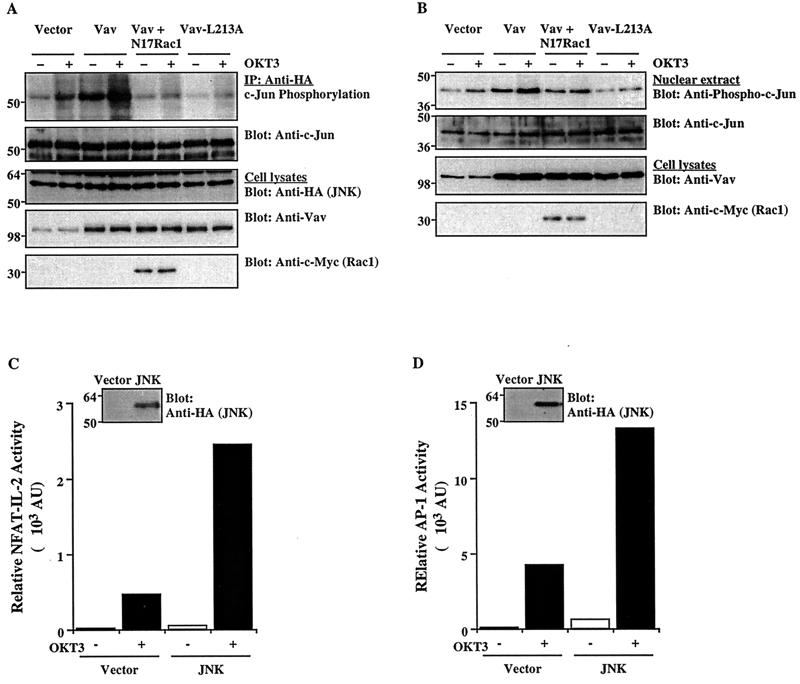
Vav, a hematopoiesis-specific signaling protein, plays an important role in T-cell development and activation. Vav upregulates the expression of the interleukin-2 (IL-2) gene, primarily via activation of the distal NFAT site in the IL-2 gene promoter (NFAT-IL-2). However, since this site cooperatively binds NFAT and AP-1, the relative contribution of Vav to NFAT versus AP-1 activation has not been determined. Here, we studied the respective roles of the AP-1 and NFAT pathways in the T-cell receptor (TCR)-mediated, Vav-dependent activation of NFAT-IL-2. Although Vav stimulated the transcriptional activity of an NFAT-IL-2 reporter gene, it failed to stimulate the transcriptional or DNA-binding activities of an AP-1-independent NFAT site derived from the human gamma interferon gene promoter. Vav also did not stimulate detectable Ca(2+) mobilization and nuclear translocation of NFATc or NFATp. On the other hand, Vav induced the activation of Rac1 or Cdc42 and c-Jun N-terminal kinase (JNK), enhanced the transcriptional and DNA-binding activities of AP-1, and induced increased phosphorylation of c-Jun. Dominant-negative Vav and/or Rac1 mutants blocked the TCR-mediated stimulation of these events, demonstrating the physiological relevance of these effects. Vav also associated with Rac1 or Cdc42 in T cells, and anti-CD3 antibody stimulation enhanced this association. These findings indicate that a Rac1-dependent JNK/c-Jun/AP-1 pathway, rather than the Ca(2+)/NFAT pathway, plays the predominant role in NFAT-IL-2 activation by Vav.


Figures

FIG. 1
FIG. 1
The Vav/Rac pathway plays a role in NFAT–IL-2 and AP-1 activation. (A) Jurkat-TAg cells were transfected with empty pEF vector (10 μg) or wild-type Vav, Vav-L213A, or Vav plus N17Rac1 (5 μg each). After 24 h, cells (107/ml) either were left unstimulated or were stimulated with cross-linked OKT3 (0.3 μg/ml). Nuclear extracts were analyzed by an EMSA using NFAT–IL-2 and AP-1 oligonucleotide probes. The membranes were subjected to autoradiography (top two panels), and total cellular extracts from the same cells were immunoblotted with anti-Vav or anti-c-Myc antibodies (bottom two panels). The results shown are representative of four separate experiments. Arrowheads indicate specific NFAT and AP-1 complexes. (B) A similar EMSA was performed using a nuclear extract from OKT3-stimulated, Vav-transfected cells in the absence (−) or presence of the indicated unlabeled competing oligonucleotides, antibodies, or control IgG. A similar inhibition pattern was observed with an extract from unstimulated, Vav-transfected cells (data not shown). (C to F) Cells were cotransfected with NFAT–IL-2–Luc (C and E) or AP-1–Luc (D and F) reporter plasmids plus the indicated combinations of Vav, Vav-L213A, Vav-6A-DH, and/or N17Rac1. After 24 h, the cells either were left unstimulated or were stimulated with the indicated concentrations of cross-linked OKT3. Luciferase and β-Gal activities in cell extracts were determined 8 h later. AU, arbitrary units. The standard deviation for triplicates was <10%, and each experiment was repeated four times, with similar results. Samples of the same lysates were analyzed for the expression of Vav and N17Rac1 by immunoblotting with anti-Vav and anti-c-Myc antibodies, respectively (insets). The positions of molecular weight standards (in thousands) are shown.
FIG. 2
FIG. 2
Vav fails to induce detectable Ca2+ mobilization or NFAT nuclear translocation. (A) Cells were transfected with empty vector, Vav, Vav-L213A, or N17Rac1. After 24 h, cells either were left unstimulated or were stimulated with cross-linked OKT3. [Ca2+]i was determined by monitoring indo-1 fluorescence. The results are representative of three experiments. Samples of the same lysates were analyzed for the expression of Vav or Rac1 by immunoblotting (insets). The positions of molecular weight standards (in thousands) are shown. (B) In a parallel experiment, the percentage of cells expressing the transfected plasmids was determined by FACS analysis of GFP-transfected cells. Based on this information, the anti-CD3 antibody-stimulated peak [Ca2+]i in the transfected cell fraction was calculated. Error bars indicate standard deviations. (C) Cells were transfected with empty vector or with Vav plasmid DNA. After 24 h, the cells either were left unstimulated or were stimulated by cross-linked OKT3. Cytosolic and nuclear extracts were immunoblotted with anti-NFATc (top panel), anti-NFATp (second panel from top) or anti-JNK (third panel from top) antibodies, as indicated. Total cellular extracts from the same groups were immunoblotted with an anti-Vav antibody (bottom panel). The results shown are representative of four separate experiments. Brackets indicate NFATc and NFATp proteins. The positions of molecular weight standards (in thousands) are shown.
FIG. 3
FIG. 3
Effects of Vav on DNA-binding and transcriptional activities of NFAT-IFN. (A) Cells were transfected with empty pEF vector, Vav, or Vav plus N17Rac1. After 24 h, cells either were left unstimulated or were stimulated with cross-linked OKT3. Nuclear extracts were prepared and analyzed by an EMSA using a 32P-labeled NFAT-IFN probe (top panel). Total cellular extracts from the same groups were immunoblotted with anti-Vav (middle panel) or anti–c-Myc (bottom panel) antibodies. The results are representative of three separate experiments. The arrowhead indicates the specific NFAT-IFN complex. (B) A similar EMSA was conducted using a nuclear extract from OKT3-stimulated, Vav-transfected cells in the absence (−) or presence of the indicated unlabeled competing oligonucleotides, antibodies, or control IgG. A similar inhibition pattern was observed with an extract from unstimulated, Vav-transfected cells (data not shown). (C) Cells were cotransfected with an NFAT-IFN–Luc reporter plasmid plus empty vector, Vav, or Vav plus N17Rac1. Stimulation and reporter activity determinations were done as described in the legend to Fig. 1. AU, arbitrary units. The data shown are representative of four experiments. Samples of the same lysates were analyzed for the expression of Vav or Rac1 by immunoblotting with anti-Vav or anti-c-Myc MAbs (insets). The positions of molecular weight standards (in thousands) are shown.
FIG. 4
FIG. 4
Interaction between Vav and Rho family GTPases. Jurkat-TAg cells were transfected with the indicated plasmids (5 μg each). After 24 h, the cells either were left unstimulated or were stimulated with cross-linked OKT3. (A) The active, GTP-loaded forms of Rac1 or Cdc42 in cell lysates were captured by incubation with a GST-PBD fusion protein, followed by immobilization on glutathione-coupled Sepharose 4B beads. Washed precipitates were analyzed by immunoblotting with the indicated antibodies (top two panels). Total cellular extracts from the same groups were immunoblotted with anti-Vav, anti-Rac1, or anti-Cdc42 antibodies (bottom three panels). The results shown are representative of five separate experiments. (B) Cell lysates were immunoprecipitated (IP) with anti-Rac1 or anti-Cdc42 antibodies or with control IgG and immunoblotted with an anti-Vav MAb. Total cellular extracts were immunoblotted as described for panel A. The results shown are representative of three separate experiments. The positions of molecular weight standards (in thousands) are shown.
FIG. 5
FIG. 5
Effect of Vav on JNK activation and c-Jun phosphorylation. (A) Cells were cotransfected with an HA-tagged JNK1 plasmid plus the indicated combinations of Vav, Vav-L213A, and/or N17Rac1 (5 μg each). After 24 h, the cells either were left unstimulated or were stimulated with cross-linked OKT3. JNK1 was immunoprecipitated (IP) from cell lysates with an anti-HA MAb and subjected to an in vitro kinase assay using GST–c-Jun as a substrate. The SDS-PAGE-separated kinase reaction was visualized by autoradiography (top panel), followed by immunoblotting with an anti-c-Jun antibody (second panel from top). Total cellular extracts were immunoblotted with anti-HA, anti-Vav, or anti-c-Myc antibodies (bottom three panels). The results shown are representative of four separate experiments. (B) Cells were transfected and stimulated as described for panel A. Nuclear extracts were prepared, resolved by SDS-PAGE, and immunoblotted with anti-phospho-c-Jun or anti-c-Jun antibodies (top two panels). Total cellular extracts from the same groups were immunoblotted with anti-Vav or anti-c-Myc antibodies (bottom two panels). (C and D) Cells were cotransfected with NFAT–IL-2–Luc or AP-1–Luc reporter plasmids plus empty pcDNA3 or an HA-tagged JNK1 plasmid. Reporter activity in unstimulated or OKT3-stimulated cells was determined as described in the legend to Fig. 1. AU, arbitrary units. Samples of the same lysates were immunoblotted with an anti-HA MAb to detect JNK expression (insets). The results are representative of three separate experiments. The positions of molecular weight standards (in thousands) are shown.

Similar articles

Cited by

References

    1. Bagrodia S, Derijard B, Davis R J, Cerione R A. Cdc42 and PAK-mediated signaling leads to Jun kinase and p38 mitogen-activated protein kinase activation. J Biol Chem. 1995;270:27995–27998. - PubMed
    1. Bagrodia S, Taylor S J, Creasy C L, Chernoff J, Cerione R A. Identification of a mouse p21Cdc42/Rac activated kinase. J Biol Chem. 1995;270:22731–22737. - PubMed
    1. Beals C R, Clipstone N A, Ho S N, Crabtree G R. Nuclear localization of NF-ATc by a calcineurin-dependent, cyclosporin-sensitive intramolecular interaction. Genes Dev. 1997;11:824–834. - PubMed
    1. Billadeau D D, Mackie S M, Schoon R A, Leibson P J. Specific subdomains of Vav differentially affect T cell and NK cell activation. J Immunol. 2000;164:3971–3981. - PubMed
    1. Chow C-W, Rincon M, Cavanagh J, Dickens M, Davis R J. Nuclear accumulation of NFAT4 opposed by the JNK signal transduction pathway. Science. 1997;278:1638–1641. - PubMed

Publication types

MeSH terms