. 2001 Apr 10;98(8):4361-6.
doi: 10.1073/pnas.071051098. Epub 2001 Apr 3.

Three-dimensional image reconstruction of dephosphorylated smooth muscle heavy meromyosin reveals asymmetry in the interaction between myosin heads and placement of subfragment 2

Affiliations

Three-dimensional image reconstruction of dephosphorylated smooth muscle heavy meromyosin reveals asymmetry in the interaction between myosin heads and placement of subfragment 2

T Wendt et al. Proc Natl Acad Sci U S A. .

Abstract

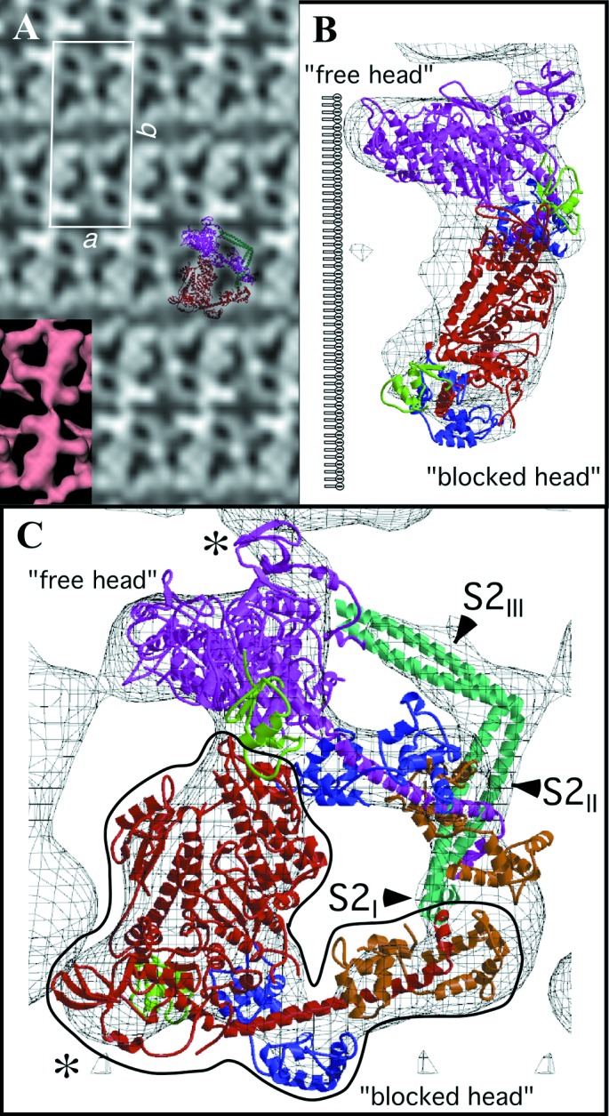
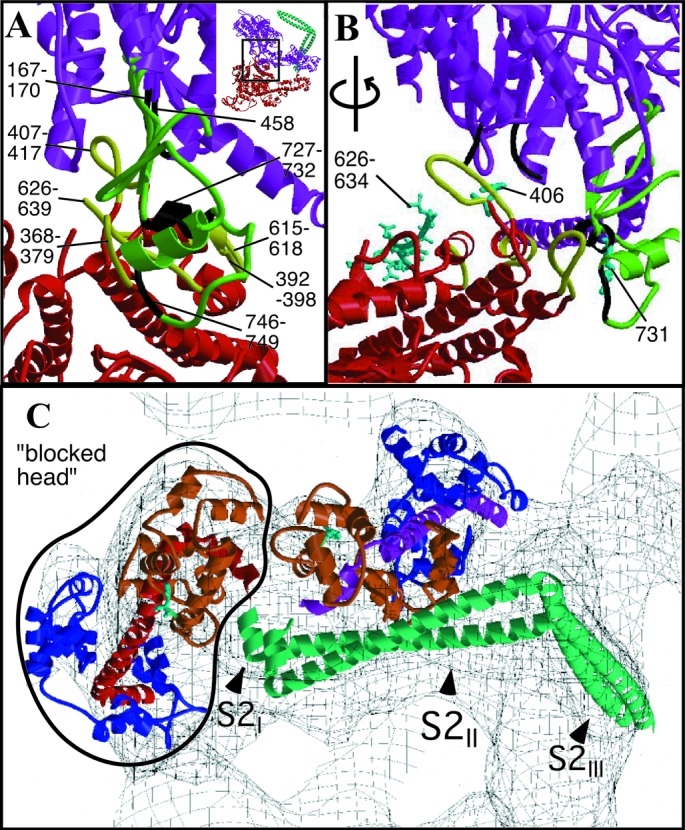
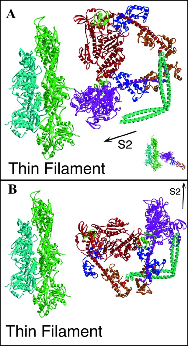
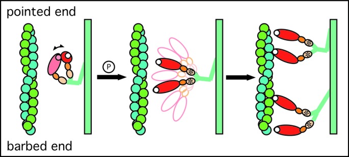
Regulation of the actin-activated ATPase of smooth muscle myosin II is known to involve an interaction between the two heads that is controlled by phosphorylation of the regulatory light chain. However, the three-dimensional structure of this inactivated form has been unknown. We have used a lipid monolayer to obtain two-dimensional crystalline arrays of the unphosphorylated inactive form of smooth muscle heavy meromyosin suitable for structural studies by electron cryomicroscopy of unstained, frozen-hydrated specimens. The three-dimensional structure reveals an asymmetric interaction between the two myosin heads. The ATPase activity of one head is sterically "blocked" because part of its actin-binding interface is positioned onto the converter domain of the second head. ATPase activity of the second head, which can bind actin, appears to be inhibited through stabilization of converter domain movements needed to release phosphate and achieve strong actin binding. When the subfragment 2 domain of heavy meromyosin is oriented as it would be in an actomyosin filament lattice, the position of the heads is very different from that needed to bind actin, suggesting an additional contribution to ATPase inhibition in situ.


Figures

Figure 1
Figure 1
Two-dimensional arrays of unphosphorylated smooth muscle HMM. (A) Averaged in-plane projection of frozen hydrated arrays with a surface view of the 3D reconstruction as Inset (Lower Left). Overlaid on the projection is a view of the 3D model reduced to the same scale. The two S1s are colored red and magenta and the S2 cyan. (B and C) Two views of the model fitted to the reconstruction. B is oriented along the crystal a axis. The location of the lipid monolayer, which is not seen in the reconstruction, is drawn to the left of the structure. C is oriented perpendicular to the plane of the crystal. The lipid monolayer would be located below the structure in this view. The “blocked” head is outlined in black. The color scheme for all ribbon diagrams is as follows: the heavy chain of the “free” myosin head is magenta, and that of the “blocked” head is red. For both myosin heads, the converter domain is green, the ELCs blue, and the RLCs orange. Figures were prepared by using bobscript and raster3d (41, 42). The region displayed in C can be seen in stereo in Fig. 5 as well as Movie 1, which are published as supplemental data on the PNAS web site, www.pnas.org.
Figure 2
Figure 2
(A and B) Heavy chain–heavy chain interactions. Color scheme as in Fig. 1, except that loops involved in heavy chain–heavy chain interactions are in black on the “free” head and in yellow on the “blocked” head. (A) View perpendicular to the crystal plane (upper right-hand corner Inset shows corresponding overall view). “Free” head residues potentially involved in the interaction include 458 (at the end of a disordered loop), loop 167–170, and converter domain loop 746–749 and helix 727–732. “Blocked” head residues include loop 368–379, the myopathy loop 407–417, loop 615–618, and helix 392–398. (B) Same region rotated by ≈70° to view within the crystal plane. Displayed in cyan in ball-and-stick format are residues 626–634 of the smooth muscle inhibitory domain (43) and “blocked” head residue 406 and “free” head residue 731. (C) S1–S2 junction viewed down the a axis of the unit cell. The light chain lever arm of the “blocked” head is outlined in black. The N termini of the RLCs (F19, smooth muscle F25) are shown in cyan as ball-and-stick structures. Figs. 6–8 are stereoviews of Fig. 2 A–C; the region shown in Fig. 2 A and B is Movie 2; Fig 2C is Movie 3. Figs. 6–8 and Movies 2 and 3 are published as supplemental data on the PNAS web site, www.pnas.org.
Figure 3
Figure 3
Docking of unphosphorylated HMM onto actin. (A) The “free” head of the HMM model is oriented on actin by using the actin-binding domain of rigor skeletal muscle actomyosin (Inset) as the reference. The smooth muscle S1 with a transition state analog at the active site appears with the lever arm up (15) in contrast to the lever arm down-postpowerstroke conformation of skeletal muscle actomyosin (35). The HMM model, when oriented via the actin-binding domain, verifies that one head can interact with actin without steric hindrance from the second head. The orientation of the double-headed myosin in B represents the arrangement with the S2lll segment oriented toward the direction of force transmission, as would occur within the muscle lattice. The head orientations in this configuration do not favor actin binding by either head.
Figure 4
Figure 4
Diagram illustrating a model for the structural changes that occur on activation of smooth muscle myosin. (A) In the “off” state, the two heads of myosin are in an orientation that is disadvantageous for actin binding by either head. (B) On phosphorylation of the RLC, the head–head interaction is interrupted, and both can search independently for actin monomers suitably placed for binding. (C) Myosin heads bind actin in the prepowerstroke position, which on filament sliding will transform to the rigor-like configuration that characterizes the end of the powerstroke. Note that the initial attachment to actin is likely to be considerably less ordered than implied by the cartoon (36).

References

    1. Sellers J R. In: Protein Profile. Sheterline P, editor. Oxford, U.K.: Oxford Univ. Press; 1999.
    1. Rayment I, Holden H M, Whittaker M, Yohn C B, Lorenz M, Holmes K C, Milligan R A. Science. 1993;261:58–65. - PubMed
    1. McLachlan A D, Karn J. J Mol Biol. 1983;164:605–626. - PubMed
    1. Sellers J R. Curr Opin Cell Biol. 1991;3:98–104. - PubMed
    1. Sellers J R. J Biol Chem. 1985;260:15815–15819. - PubMed

Publication types

Substances

Associated data

LinkOut - more resources