. 2001 Apr 10;98(8):4337-42.
doi: 10.1073/pnas.071514398. Epub 2001 Apr 3.

Crystal structure of cis-prenyl chain elongating enzyme, undecaprenyl diphosphate synthase

Affiliations

Crystal structure of cis-prenyl chain elongating enzyme, undecaprenyl diphosphate synthase

M Fujihashi et al. Proc Natl Acad Sci U S A. .

Abstract

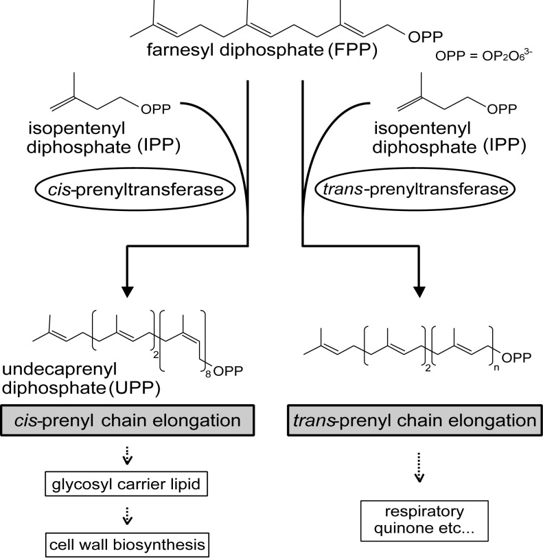
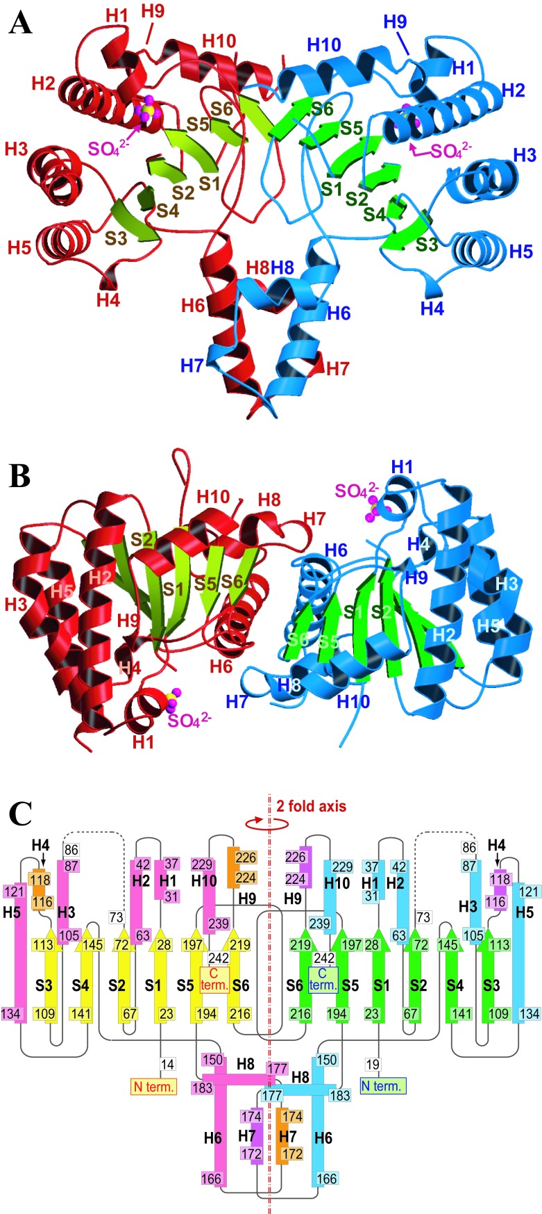
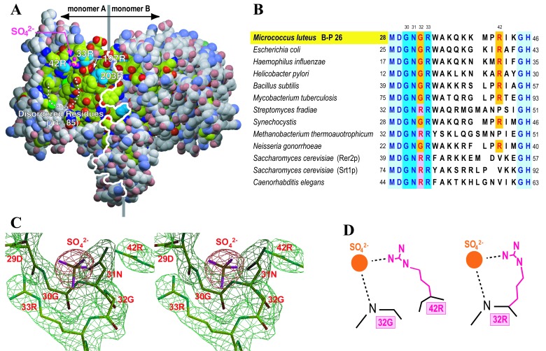
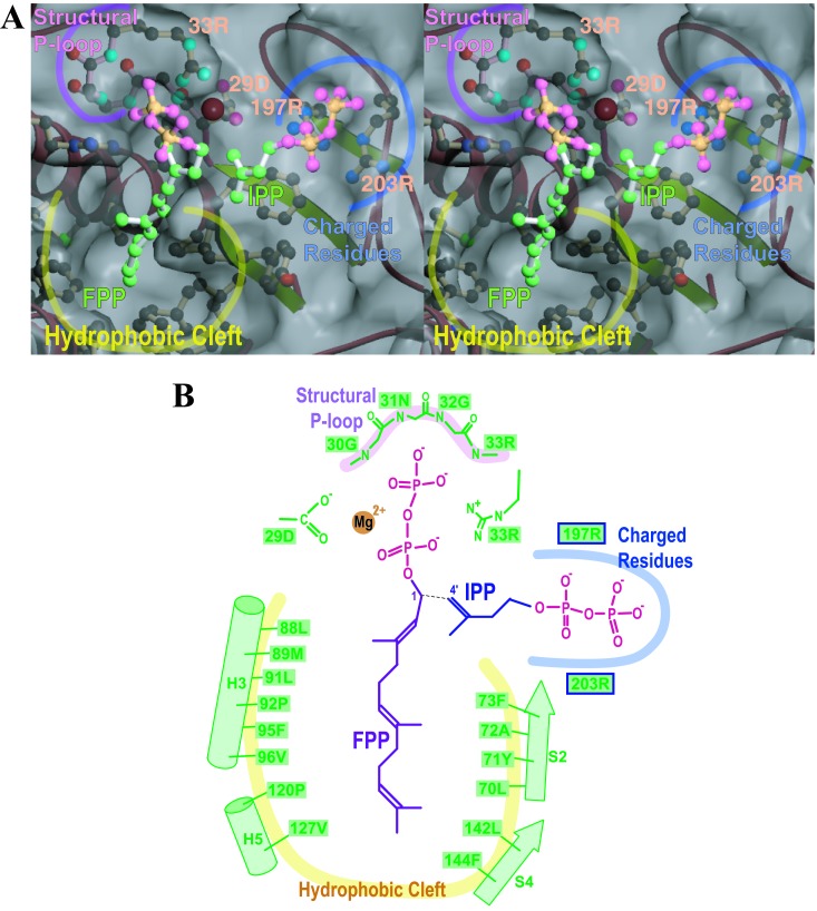
Undecaprenyl diphosphate synthase (UPS) catalyzes the cis-prenyl chain elongation onto trans, trans-farnesyl diphosphate (FPP) to produce undecaprenyl diphosphate (UPP), which is indispensable for the biosynthesis of bacterial cell walls. We report here the crystal structure of UPS as the only three-dimensional structure among cis-prenyl chain elongating enzymes. The structure is classified into a protein fold family and is completely different from the so-called "isoprenoid synthase fold" that is believed to be a common structure for the enzymes relating to isoprenoid biosynthesis. Conserved amino acid residues among cis-prenyl chain elongating enzymes are located around a large hydrophobic cleft in the UPS structure. A structural P-loop motif, which frequently appears in the various kinds of phosphate binding site, is found at the entrance of this cleft. The catalytic site is determined on the basis of these structural features, from which a possible reaction mechanism is proposed.


Figures

Figure 1
Figure 1
A schematic drawing of the biosynthesis related to prenyl chains. UPS catalyzes 8× cis-prenyl chain elongation step by step. Both cis- and trans-prenyl chains are constructed from the same substrates by the cis or trans types of prenyltransferase, in which the only difference is the cis- and trans-isomerism of the prenyl chain product.
Figure 2
Figure 2
Overall structure of UPS from M. luteus B-P 26. A front view (A) and a top view (B) of the dimer structure (ribbon model). One monomer of UPS is shown by red helices (α- and 310-helices) and yellowish green arrows (β-strands), and the other monomer by blue helices and green arrows. Helices (H1–H10) and strands (S1–S6) are labeled together with the sulfate ions found in the crystal structure. These two figures were prepared with molscript (33) and raster3d (34). (C) A topology diagram of the secondary structure of the UPS dimer. α- and 310-helices of one monomer and those of the other monomer are colored pink, orange, sky-blue, and light purple, respectively. β-strands of one monomer and the other are colored yellow and green, respectively.
Figure 3
Figure 3
Active site in the UPS structure. (A) Space filling model of the UPS dimer. C atoms are indicated in yellowish green and gray, in which the former color corresponds to conserved residues and the latter nonconserved residues among the cis-prenyltransferase family. These conserved residues form a large hydrophobic cleft. N, O, and S atoms are colored in blue, red, and green, respectively. The white line indicates the interface of the dimer, where the left and right monomers are named A and B. White dotted line shows the disordered residues from 74S to 85V. This figure was prepared in the same manner as Fig. 2 A and B. (B) A comparison of amino acid sequences in 13 proteins that have sequence homology with UPS from M. luteus B-P 26 (12, 13). Only residues corresponding to 28–46 of M. luteus UPS are presented. The proteins from M. luteus, E. coli, and Hemophilus influenzae demonstrate UPS activity (10, 12, 13). RER2p from S. cerevisiae has cis-prenyltransferase activity (11). The others are potential cis-prenyltransferases homologous with M. luteus UPS. (C) Stereo view of the electron density map around the structural P-loop motif and the sulfate ion. The final refined structure is superimposed. (D) Three-dimensionally conserved guanidinium head of arginine. The positively charged guanidinium group of either 42R (in the case of 32G; Left) or 32R (in the case of 32R; Right) binds to the diphosphate head of the substrate.
Figure 4
Figure 4
Hypothetical FPP and IPP binding model with UPS. (A) Stereo view of the molecular surface of UPS (gray envelope) and the bindings of FPP and IPP (C, O, and P atoms in green, pink, and orange, respectively). The estimated position of the magnesium ion is shown by the brown sphere. This figure was prepared by using molscript (33), grasp (35), and raster3d (34). (B) Schematic presentation of a binding model. The UPS structure is shown in green.

References

    1. Ogura K, Koyama T. Chem Rev. 1998;98:1263–1276. - PubMed
    1. Carattoli A, Romano N, Ballario P, Morelli G, Macino G. J Biol Chem. 1991;266:5854–5859. - PubMed
    1. Joly A, Edwards P A. J Biol Chem. 1993;268:26983–26989. - PubMed
    1. Koyama T, Obata S, Osabe M, Takeshita A, Yokoyama K, Uchida M, Nishino T, Ogura K. J Biochem (Tokyo) 1993;113:355–363. - PubMed
    1. Song L, Poulter C D. Proc Natl Acad Sci USA. 1994;91:3044–3048. - PMC - PubMed

Publication types

Substances

Associated data

LinkOut - more resources