

Power Doppler sonography to differentiate tuberculous cervical lymphadenopathy from nasopharyngeal carcinoma
- PMID: 11290489
- PMCID: PMC7976010
Power Doppler sonography to differentiate tuberculous cervical lymphadenopathy from nasopharyngeal carcinoma
Abstract
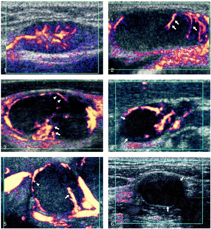
Background and purpose: Tuberculous lymphadenitis and metastatic nodes from nasopharyngeal carcinoma are common in Asians and are often indistinguishable clinically. Because their treatment depends on prompt diagnosis, we undertook this study to evaluate if power Doppler sonography could distinguish these two pathologic abnormalities. The intranodal vascular appearances of tuberculous neck nodes are compared with benign reactive neck nodes and metastatic nodes from nasopharyngeal carcinoma.
Methods: The appearances of power Doppler sonograms of 42 tuberculous nodes were compared with 28 metastatic nodes from nasopharyngeal carcinoma and 27 benign reactive nodes. The intranodal distribution of vessels and the intranodal vascular resistance of vessels were compared among these three groups. All examinations were performed by the same sonologist (A.A.), who had more than 3 years' scanning experience, and all data analysis was performed by the same investigator (M.Y.).
Results: The intranodal vascular distribution in tuberculous nodes was varied and simulated both benign and malignant disease. Avascularity of nodes and displacement of hilar vascularity were frequent in tuberculous nodes. Metastatic nodes from nasopharyngeal carcinoma (resistive index [RI], 0.81+/-0.09; pulsatile index [PI], 1.91+/-0.81) had a higher vascular resistance than did tuberculous nodes (RI, 0.71+/-0.11; PI, 1.34+/-0.55). Tuberculous nodes had a higher vascular resistance than did reactive nodes (RI, 0.66+/-0.09; PI, 1.10+/-0.26).
Conclusion: Avascularity, displaced hilar vessels, and low intranodal vascular resistance are clues that may suggest the tuberculous nature of neck nodes. However, there is overlap of appearance between tuberculous nodes, benign reactive neck nodes, and metastatic nodes. Thus, histologic analysis is often required for a definitive diagnosis.
Figures
References
-
- Ahuja A, Ying M, King W, Metreweli C. A practical approach to ultrasound of cervical lymph nodes. J Laryngol Otol 1997;111:245-256 - PubMed
-
- Ying M, Ahuja AT, Evans R, King W, Metreweli C. Cervical lymphadenopathy: sonographic differentiation between tuberculous nodes and nodal metastases from non-head and neck carcinomas. J Clin Ultrasound 1998;26:383-389 - PubMed
-
- Baatenburg de Jong RJ, Rongen RJ, Verwoerd CD, van Overhagen H, Lameris JS, Knegt P. Ultrasound-guided fine-needle aspiration biopsy of neck nodes. Arch Otolaryngol Head Neck Surg 1991;117:402-404 - PubMed
-
- Steinkamp HJ, Meuffelmann M, Bock JC, Thiel T, Kenzel P, Felix R. Differential diagnosis of lymph node lesions: a semiquantitative approach with colour Doppler ultrasound. Br J Radiol 1998;71:828-833 - PubMed
Publication types
MeSH terms
LinkOut - more resources
Full Text Sources
Research Materials
Miscellaneous