

Global estimation of myelination in the developing brain on the basis of magnetization transfer imaging: a preliminary study
- PMID: 11290496
- PMCID: PMC7976006
Global estimation of myelination in the developing brain on the basis of magnetization transfer imaging: a preliminary study
Abstract
Background and purpose: In the developing brain, myelination occurs in an orderly and predetermined sequence. The aim of this study was to determine whether such changes can be tracked using volumetric magnetization transfer imaging.
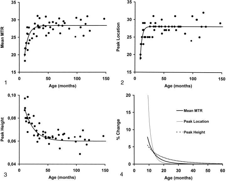
Methods: Three-dimensional magnetization transfer imaging was performed in 50 children (age range, 0.6-190 months) with no evidence of developmental delay or structural abnormalities. Volumetric magnetization transfer ratio (MTR) parameters generated of the whole brain were mean MTR and height and location of the MTR histogram peak. Relationships between volumetric MTR parameters and age were assessed using nonlinear regression analysis.
Results: With age, all volumetric MTR parameters changed exponentially in a way that was best expressed by the function y = a + b.exp(-x/c) (P < .0001). The peak height of the MTR histogram was the parameter that changed most predictably and that continued to change for the longest period of time.
Conclusion: With this preliminary study, we show that by using volumetric MTR analysis, it is possible to monitor changes in the developing brain, presumably the myelination progress. This method has a potential role for detecting myelination disorders in the pediatric population, for studying the natural history of these diseases, and for monitoring the effects of treatment.
Figures
References
-
- McArdle CB, Richardson CJ, Nicholas DA, Mirfakhraee M, Hayden CK, Amparo EG. Developmental features of the neonatal brain: MR imaging: part I. gray-white matter differentiation and myelination. Radiology 1987;162:223-229 - PubMed
-
- Barkovich AJ, Kjos BO, Jackson DE Jr, Norman D. Normal brain maturation of the neonatal and infant brain: MR imaging at 1.5 T. Radiology 1988;166:173-180 - PubMed
-
- Dietrich RB, Bradley WG. Normal and abnormal white matter maturation. Semin Ultrasound CT MR 1988;9:192-200 - PubMed
MeSH terms
LinkOut - more resources
Full Text Sources
Medical